. 2008 May 22;453(7194):519-23.
doi: 10.1038/nature06968.

The ground state of embryonic stem cell self-renewal

Affiliations

The ground state of embryonic stem cell self-renewal

Qi-Long Ying et al. Nature. .

Abstract

In the three decades since pluripotent mouse embryonic stem (ES) cells were first described they have been derived and maintained by using various empirical combinations of feeder cells, conditioned media, cytokines, growth factors, hormones, fetal calf serum, and serum extracts. Consequently ES-cell self-renewal is generally considered to be dependent on multifactorial stimulation of dedicated transcriptional circuitries, pre-eminent among which is the activation of STAT3 by cytokines (ref. 8). Here we show, however, that extrinsic stimuli are dispensable for the derivation, propagation and pluripotency of ES cells. Self-renewal is enabled by the elimination of differentiation-inducing signalling from mitogen-activated protein kinase. Additional inhibition of glycogen synthase kinase 3 consolidates biosynthetic capacity and suppresses residual differentiation. Complete bypass of cytokine signalling is confirmed by isolating ES cells genetically devoid of STAT3. These findings reveal that ES cells have an innate programme for self-replication that does not require extrinsic instruction. This property may account for their latent tumorigenicity. The delineation of minimal requirements for self-renewal now provides a defined platform for the precise description and dissection of the pluripotent state.


Figures

Figure 1
Figure 1. Three inhibitors (3i) support robust self-renewal and de novo derivation of pluripotent ES cells
a, Immunostaining of E14Tg2a ES cells with Oct4 after four passages in N2B27 plus PD184352 and SU5402 (PS). b, RT–PCR analysis of marker expression in ES cells in N2B27 containing PS with or without LIF. Gapdh, gene encoding glyceraldehyde-3-phosphate dehydrogenase. c, Low-magnification phase-contrast image of ES cells passaged in N2B27 plus CHIR99021 showing a mixture of differentiated cells with compact undifferentiated colonies (arrowheads). d, Immunostaining with Oct4 after several passages in N2B27 plus 3i, showing compact colony morphology. e, RT–PCR analysis of marker expression in ES cells cultured in N2B27 alone (−) or with LIF and BMP4 (LB) or 3i. f, Phase and fluorescence images of expansion from a single Oct4GiP ES cell in 3i. g, Cloning efficiencies of E14Tg2a ES cells after single-cell deposition in the indicated conditions (top), and in CHIR99021 plus PD0325901 (2i, see Fig. 2) with or without B27, or with the replacement of serum albumin with recombinant albumin (rHSA) (bottom; experiment performed in 5% O2). h, Oct4GIP ES cells cultured for five passages (total 28 days) in basal medium supplemented with transferrin and BSA only plus 3 μM CHIR99021, 0.5 μM PD184352 and 1 μM SU5402. i, Chimaera and germline offspring produced from CBA ES cells derived in 3i. Chimaera showing extensive contribution of CBA (agouti coat colour) ES cells mated with C57BL/6 (black) produced agouti pups, indicating the transmission of the CBA genome.
Figure 2
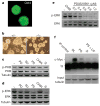
Figure 2. Effects of 3i components on intracellular signalling cascades
a, E14Tg2a ES cells remain undifferentiated and Oct4-positive in alternative 3i with SU5402 replaced by PD173074. b, fgf4-null ES cells expand without differentiation in N2B27 plus CHIR99021 only, without a requirement for FGFR/MEK inhibition. c, d, Immunoblot analyses of steady-state levels of phospho(Ser 473)-PKB (p-PKB) (c) and phospho(Thr 202, Tyr 204)-ERK (p-ERK) (d) in ES cells after 24 h in N2B27 alone (−), plus 0.8 μM PD184352 (PD), 2 μM SU5402 (SU), 3 μM CHIR99021, PS or 3i. e, Immunoblot analyses of phospho(Thr 202, Tyr 204)-ERK levels in ES cells after 24 h in N2B27 alone (−), plus 3 μM CHIR99021 (CHIR) or 3 μM CHIR99021 plus PD0325901 at the indicated concentrations. f, c-Myc protein in ES cells assayed by sequential immunoprecipitation (IP) and immunoblotting after 24 h in serum plus LIF (GL), PS, 3i, or PS plus LIF. IP control is the GL sample immunoprecipitated with anti-tubulin. Input samples were subjected to SDS PAGE and blotted for tubulin to control for loading.
Figure 3
Figure 3. ES-cell propagation in 3i does not involve STAT3
a, Top: genomic PCR for null and wild-type (WT) Stat3 alleles in heterozygous and Stat3 homozygous null ES cells. Bottom: immunoblot analysis of heterozygous and Stat3 homozygous null ES cells. E14, E74Tg2a ES cells. b, Oct4 immunostaining of Stat3-null ES cells. c, Quantitative RT–PCR analysis of undifferentiated Stat3-null ES cells and derivative embryoid bodies (EB) at days 3 and 6. d, Stat3-null ES cells generate morphologically differentiated cells expressing the neuronal marker βIII-tubulin (TuJ1 immunoreactive). e, Quantitative RT–PCR analysis of Socs3 (Stat3 target gene; filled columns) and Egr1 (ERK target gene; open columns) expression in undifferentiated Stat3 wild-type and null ES cells grown in N2B27 alone or stimulated with LIF for 1 h. f, Stat3-null MF1 ES cells differentiate in the presence of LIF and feeders, in contrast with wild-type MF1 ES cells, which remain undifferentiated.
Figure 4
Figure 4. CHIR99021 acts via inhibition of GSK3 to enhance ES-cell growth capacity and viability
a, TOPFlash assay in ΔNLef1 stable transfectants. Results are shown as means and s.d. for three biological replicates. Non, N2B27 alone; CHIR, CHIR99021. b, Competitive growth assay for three clones of ΔNLef1 stable transfectants and Cre revertants (GFP+) in serum and LIF or in 3i. Results are shown as means and s.d. for four biological replicates. c, GSK3α/β-deficient DKO cells cultured in the indicated conditions for two passages. L, LIF. d, Phase-contrast and Oct4 immunostaining of DKO cells after eight passages in 1 μM PD0325901. Parallel cultures with addition of CHIR99021 were indistinguishable. e, ES cells constitutively expressing Nanog respond to CHIR99021 by enhanced self-renewal at low density in 3i compared with PS. f, ES cells self-renew in 2 μM PD0325901 plus CHIR99021. g, h, Diagrams of self-replication of the pluripotent state when inductive phospho-ERK signalling is either inhibited upstream by chemical antagonists (g) or counteracted downstream by LIF and BMP (h). Inhibition of GSK3 serves a key function in augmenting self-renewal when phospho-ERK (pERK) is suppressed by maintaining cellular growth capacity and additionally reinforcing suppression of neural commitment.

References

    1. Martin GR. Isolation of a pluripotent cell line from early mouse embryos cultured in medium conditioned by teratocarcinoma stem cells. Proc Natl Acad Sci USA. 1981;78:7634–7638. - PMC - PubMed
    1. Evans MJ, Kaufman M. Establishment in culture of pluripotential cells from mouse embryos. Nature. 1981;292:154–156. - PubMed
    1. Rathjen J, et al. Formation of a primitive ectoderm like cell population, EPL cells, from ES cells in response to biologically derived factors. J Cell Sci. 1999;112:601–612. - PubMed
    1. Sato N, Meijer L, Skaltsounis L, Greengard P, Brivanlou AH. Maintenance of pluripotency in human and mouse embryonic stem cells through activation of Wnt signaling by a pharmacological GSK-3-specific inhibitor. Nature Med. 2004;10:55–63. - PubMed
    1. Ying QL, Nichols J, Chambers I, Smith A. BMP induction of Id proteins suppresses differentiation and sustains embryonic stem cell self-renewal in collaboration with STAT3. Cell. 2003;115:281–292. - PubMed

Publication types

MeSH terms