. 2008 May 22;453(7194):548-52.
doi: 10.1038/nature06924.

Ubiquitin docking at the proteasome through a novel pleckstrin-homology domain interaction

Affiliations

Ubiquitin docking at the proteasome through a novel pleckstrin-homology domain interaction

Patrick Schreiner et al. Nature. .

Abstract

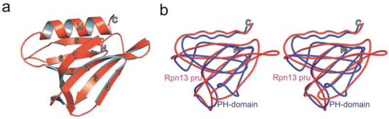
Targeted protein degradation is largely performed by the ubiquitin-proteasome pathway, in which substrate proteins are marked by covalently attached ubiquitin chains that mediate recognition by the proteasome. It is currently unclear how the proteasome recognizes its substrates, as the only established ubiquitin receptor intrinsic to the proteasome is Rpn10/S5a (ref. 1), which is not essential for ubiquitin-mediated protein degradation in budding yeast. In the accompanying manuscript we report that Rpn13 (refs 3-7), a component of the nine-subunit proteasome base, functions as a ubiquitin receptor, complementing its known role in docking de-ubiquitinating enzyme Uch37/UCHL5 (refs 4-6) to the proteasome. Here we merge crystallography and NMR data to describe the ubiquitin-binding mechanism of Rpn13. We determine the structure of Rpn13 alone and complexed with ubiquitin. The co-complex reveals a novel ubiquitin-binding mode in which loops rather than secondary structural elements are used to capture ubiquitin. Further support for the role of Rpn13 as a proteasomal ubiquitin receptor is demonstrated by its ability to bind ubiquitin and proteasome subunit Rpn2/S1 simultaneously. Finally, we provide a model structure of Rpn13 complexed to diubiquitin, which provides insights into how Rpn13 as a ubiquitin receptor is coupled to substrate deubiquitination by Uch37.


Figures

Figure 1
Figure 1. Crystal structure of mRpn13 Pru reveals typical Pleckstrin homology (PH) fold
a, Ribbon representation of Rpn13 Pleckstrin-like receptor for ubiquitin (Rpn13 Pru). The PH fold consisting of a seven-stranded β-sandwich structure (1–7) capped by the C-terminal α-helix. b, Stereo representation of the structural alignment of Rpn13 Pru (red) and the PH domain (blue) from Pleckstrin (PDB accession code 1PLS).
Figure 2
Figure 2. Structure of Rpn13 Pru:ubiquitin complex defines a novel ubiquitin-binding motif
a, Representative NOE interactions identified between Rpn13 Pru and ubiquitin. Each panel contains a selected region of an 15N dispersed NOESY experiment recorded on 15N, 13C and 70% 2H-labeled hRpn13 Pru mixed with equimolar quantities of unlabeled ubiquitin. All of the resonances displayed in this panel were unambiguously assigned as intermolecular NOE interactions with ubiquitin. Ubiquitin and Rpn13 Pru assignments are provided at the top and bottom of the expanded regions, respectively. b, Stereo representation of the mRpn13 Pru:ubiquitin complex oriented with ubiquitin at the top. At the interaction surface, secondary structural elements of ubiquitin and Rpn13 Pru are displayed in green and blue, respectively. Residues at the contact surface with intermolecular NOEs are in yellow (ubiquitin) or red (Rpn13 Pru) whereas those suggested to be at the contact surface only by the NMR titration experiments are displayed in purple (ubiquitin) or cyan (Rpn13 Pru). M31, C88, and E111, which shift upon hRpn2 (797–953) addition are displayed in dark green. c, Specific amino acid substitutions were made within the S2–S3, S4–S5, and S6–S7 loops of mRpn13 Pru by in vitro mutagenesis and the protein products expressed as GST-fusions and used in GST pull down assays to highlight the importance of these loops for tetraubiquitin binding. d, Ubiquitin (blue), hRpn2 (797–953) (green), or ubiquitin and hRpn2 (797–953) (red) were added to hRpn13 Pru domain, which was monitored by 1H, 15N HSQC experiments. Comparison with the spectrum acquired on the protein alone (black) indicates that S55, F76 and D78 bind ubiquitin in a manner that is independent of hRpn2 (797–953), whereas M31, C88 and E111 bind hRpn2 in a ubiquitin -independent manner.
Figure 3
Figure 3
a, Comparison of spectra acquired with monoubiquitin versus K48-linked tetraubiquitin reveal identical affects for Rpn13’s L56, F76 and F98 but differences for L73 and R104. Monoubiquitin (red) or tetraubiquitin (cyan) was added to hRpn13, which was monitored by 1H, 15N HSQC experiments. The spectrum of free hRpn13 is indicated in black, and the molar ratio of monoubiquitin (red) or tetraubiquitin (cyan) to hRpn13 was 1:1 in the represented spectra. b, Computer-generated model of the mRpn13 Pru:diubiquitin complex. White and grey ribbon diagrams display the proximal and distal ubiquitin, respectively, whereas a balls-and-sticks representation is used for the K48–G76 isopeptide bond linkage. Rpn13 Pru is colored in yellow and loops recognizing ubiquitin in blue, whereas L73, K103 and R104 are displayed in red. Diubiquitin was created by Insight II software based on atomic coordinates for the mRpn13 Pru:ubiquitin complex and monoubiquitin (PDB entry 1D3Z). In this model, the distal subunit of diubiquitin is positioned arbitrary, as its only constraints prohibit steric clashes with other atoms. c, hRpn13 Pru interacts with the I44 δ1 and A46 methyl groups of the proximal subunit. 1H, 13C HMQC spectra were acquired on samples containing either no (black) or equimolar unlabeled hRpn13 (1–150) (red) mixed with diubiquitin in which its proximal (left) or distal (right) subunit is 13C labeled. The shifted resonances are labeled. d, Model for how Rpn13 participates in Uch37 deconjugation of ubiquitinated substrates. Rpn13’s C-terminal domain (grey) binds Uch37 (orange) as its N-terminal domain binds the polyubiquitin chain and Rpn2/S1 (yellow). In this model, Uch37 binds to the distal subunit (light blue) of the chain while Rpn13 binds the proximal subunit (dark blue).
Figure 4
Figure 4. Structural comparison of ubiquitin receptors complexed with ubiquitin
a–f, Complex structures of ubiquitin and specific receptors displayed with ubiquitin in the same orientation (grey) and ubiquitin’s I44 shown in black sticks. Each receptor has unique color coding: a, Rpn13 Pru (red); b, Cue2–1CUE (PDB code 1OTR; light green); c, Dsk2UBA (PDB code 1WR1; dark green); d, S5a-UIM1 (PDB code 1YX5; light blue); e, Rabex-5 MIU/IUIM (PDB code 2FIF; dark blue); f, EAP45 GLUE domain (PDB code 2HTH; yellow). All structures are compared by a best-fit superposition of bound ubiquitin (grey). In c, L1 denotes the loop connecting α1 and α2.

Comment in

References

    1. Deveraux Q, Ustrell V, Pickart C, Rechsteiner M. A 26 S protease subunit that binds ubiquitin conjugates. J Biol Chem. 1994;269:7059–61. - PubMed
    1. Fu H, et al. Multiubiquitin chain binding and protein degradation are mediated by distinct domains within the 26 S proteasome subunit Mcb1. J Biol Chem. 1998;273:1970–81. - PubMed
    1. Jorgensen JP, et al. Adrm1, a putative cell adhesion regulating protein, is a novel proteasome-associated factor. J Mol Biol. 2006;360:1043–52. - PubMed
    1. Yao T, et al. Proteasome recruitment and activation of the Uch37 deubiquitinating enzyme by Adrm1. Nat Cell Biol. 2006;8:994–1002. - PubMed
    1. Hamazaki J, et al. A novel proteasome interacting protein recruits the deubiquitinating enzyme UCH37 to 26S proteasomes. Embo J. 2006;25:4524–36. - PMC - PubMed

Publication types

MeSH terms