. 2008 May 23;4(5):e1000071.
doi: 10.1371/journal.ppat.1000071.

Role for A-type lamins in herpesviral DNA targeting and heterochromatin modulation

Affiliations

Role for A-type lamins in herpesviral DNA targeting and heterochromatin modulation

Lindsey Silva et al. PLoS Pathog. .

Abstract

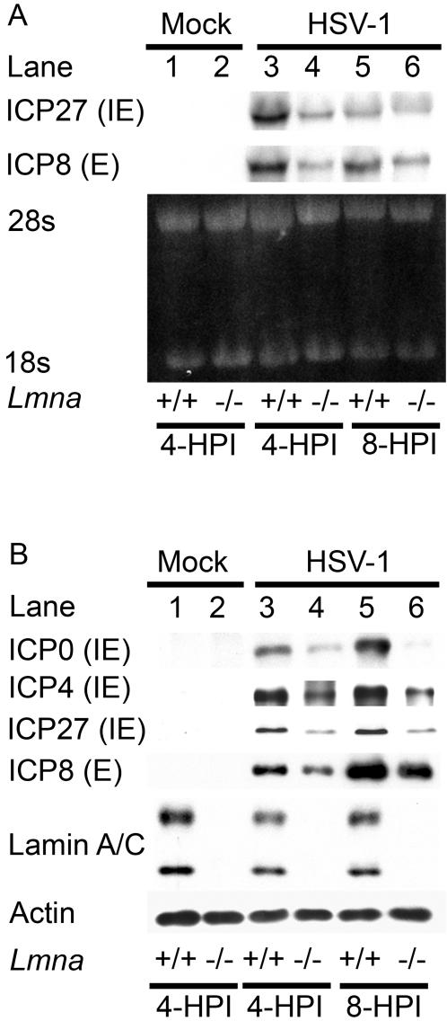
Posttranslational modification of histones is known to regulate chromatin structure and transcriptional activity, and the nuclear lamina is thought to serve as a site for heterochromatin maintenance and transcriptional silencing. In this report, we show that the nuclear lamina can also play a role in the downregulation of heterochromatin and in gene activation. Herpes simplex virus DNA initiates replication in replication compartments near the inner edge of the nucleus, and histones are excluded from these structures. To define the role of nuclear lamins in HSV replication, we examined HSV infection in wild-type and A-type lamin-deficient (Lmna-/-) murine embryonic fibroblasts (MEFs). In Lmna-/- cells, viral replication compartments are reduced in size and fail to target to the nuclear periphery, as observed in WT cells. Chromatin immunoprecipitation and immunofluorescence studies demonstrate that HSV DNA is associated with increased heterochromatin in Lmna-/- MEFs. These results argue for a functional role for A-type lamins as viral gene expression, DNA replication, and growth are reduced in Lmna-/- MEFs, with the greatest effect on viral replication at low multiplicity of infection. Thus, lamin A/C is required for targeting of the viral genome and the reduction of heterochromatin on viral promoters during lytic infection. The nuclear lamina can serve as a molecular scaffold for DNA genomes and the protein complexes that regulate both euchromatin and heterochromatin histone modifications.


Conflict of interest statement

The authors have declared that no competing interests exist.

Figures

Figure 1
Figure 1. Replication compartments are reduced in size and histone H1 is not excluded from replication compartments in Lmna−/− MEFs.
Lmna+/+ (panels a, b, e, f, I, and j) and Lmna −/− (panels c, d, g, h, k and l) MEFs were either mock-infected or infected with HSV and fixed at 8 hpi (panel A) or 4 hpi (panel B). Fixed cells were processed for indirect immunofluorescence with antibodies specific for histone H1 (green) and either HSV-1 ICP8 (panel A; red) or ICP4 (panel B; red). Scale bars equal 5 µm.
Figure 2
Figure 2. ICP4/8 foci fail to develop asymmetrically along the interior of the nuclear envelope in Lmna−/− cells.
Lmna+/+ and Lmna−/− MEFs were infected at 0.005 PFU/cell with HSV and fixed at 36 hpi following the onset of plaque formation. Cells were co-stained with ICP4 antibody (red), ICP8 antibody (green), and DAPI (blue). A) Example of Lmna +/+ MEF showing asymmetric distribution of ICP4/ICP8 foci. B) Example of Lmna −/− MEF showing symmetric distribution of ICP4/ICP8 foci. C) Quantification of ICP4/ICP8 distribution patterns in Lmna +/+ and Lmna −/− cells around plaques (n = 100 cells; 3 experiments).
Figure 3
Figure 3. HSV immediate-early (IE) and early (E) gene expression is decreased in the absence of lamin A/C.
Lmna+/+ and Lmna−/− MEFs were mock-infected (lanes 1 and 2) or infected with HSV-1 at an MOI of 20 for 4 (lanes 3 and 4) or 8 hpi (lanes 5 and 6). At the times indicated, the cells were harvested RNA and protein analysis. A) Northern blot hybridization. Equivalent amounts of total RNA were resolved in a denaturing formaldehyde agarose gel stained with ethidium bromide. The mRNAs encoding ICP27 or ICP8 were detected by Northern blot hybridization using 32P-labeled DNA. B) Western blot analysis was performed with antibodies specific for the IE proteins ICP0, ICP4, and ICP27 and the E protein ICP8. rRNAs and actin were used as loading controls for the Northern and western blots, respectively.
Figure 4
Figure 4. Heterochromatin exclusion from replication compartments during HSV-1 infection requires lamin A/C.
Lmna+/+ and Lmna−/− MEFs were either mock-infected or infected with HSV-1 at an MO of 20, fixed at 8 hpi, and stained with antibodies against ICP8 (green) and either trimethyl H3K9 (Panel A; red), trimethyl H4K20 (panel B; red), or heterochromatin protein 1α (panel C; red).
Figure 5
Figure 5. Heterochromatin on HSV-1 lytic promoter in the absence of lamin A/C.
ChIP assays were performed using antibodies specific for histone H3, trimethyl H3K9, trimethyl H3K27, or trimethyl H4K20 with lysates prepared from HSV infected Lmna+/+ and Lmna−/− MEFs at 4 hpi. The amount of ICP4 promoter sequence immunoprecipitated with each antibody was determined as described in the Materials and Methods and presented as fold enrichment relative to the cellular GAPDH gene. The data shown are the mean with error bars representing the standard error of the mean for three experiments.
Figure 6
Figure 6. Restoration of wild-type cell phenotype by expression of lamin A in Lmna−/− MEFs.
Lmna −/− MEFs were transfected with plasmids encoding either GFP (a–e) or GFP-lamin A (f–j). At 48 hours following transfection, cells were infected with HSV-1 at a MOI of 20 and processed for indirect immunofluorescence at 8 hpi. Antibodies for trimethyl H3K9 (b,g; blue) and ICP8 (c,h; red) were used. Double merged images of trimethyl H3K9 and ICP8 are shown in panels d and i. Triple merged images of GFP/GFP-lamin A (green), trimethyl H3K9 and ICP8 are shown in panels e and j. Scale bar = 5 µm.
Figure 7
Figure 7. Viral DNA replication and growth defects in the absence of lamin A/C.
A) Kinetics of viral DNA replication in Lmna+/+ and Lmna−/− cells. DNA was extracted from whole cell lysate of mock- or HSV-infected Lmna+/+ and Lmna−/− MEFs at the times indicated and quantified by real-time PCR. Relative viral DNA levels, as calculated in Materials and Methods, are shown. The error bars represent the standard deviation from three experiments. B) HSV growth curve at high MOI. Lmna+/+ and Lmna−/− MEFs were infected with HSV at an MOI of 10. At the times indicated, cells and supernatant were harvested, and total infectious virus was measured by plaque assay on Vero cells. The results shown were from triplicate cultures. C) HSV growth curve at low MOI. Lmna+/+ and Lmna−/− MEFs were infected with HSV at an MOI of 0.1. At the times indicated, cells and supernatant were harvested and titered on Vero cells, as described above. The results shown were from triplicate cultures.
Figure 8
Figure 8. Replication compartment formation and heterochromatin distribution at low multiplicity of infection.
Lmna+/+ (a–f) and Lmna−/− (g–l) MEFs were infected with HSV-1 at an MOI of 0.1 and fixed at 8 hpi (a–c, g–i) or 12 hpi (d–f, j–l). Fixed cells were processed for indirect immunofluorescence using antibodies specific for ICP8 (b, e, h, k; green) and trimethyl H3K9 (a, d, g, j; red). Merged images are shown in the bottom row. Scale bar = 5 µm.

References

    1. Roizman B, Knipe DM, Whitley RJ. Herpes Simplex Virus. In: Knipe DM, Howley PM, editors. Fields Virology. 5th ed. Philadelphia: Lippincott, Williams and Wilkins; 2007. pp. 2501–2602.
    1. Honess RW, Roizman B. Regulation of herpesvirus macro-molecular synthesis. I. Cascade regulation of the synthesis of three groups of viral proteins. J Virol. 1974;14:8–19. - PMC - PubMed
    1. Quinlan MP, Chen LB, Knipe DM. The intranuclear location of a herpes simplex virus DNA-binding protein is determined by the status of viral DNA replication. Cell. 1984;36:857–868. - PubMed
    1. de Bruyn Kops A, Knipe DM. Formation of DNA replication structures in herpes virus-infected cells requires a viral DNA binding protein. Cell. 1988;55:857–868. - PubMed
    1. Taylor TJ. Intranuclear localization of the herpes simplex virus ICP8 protein [Dissertation] Cambridge, MA: Harvard University; 2002.

Publication types

MeSH terms