. 2008 May 23;4(5):e1000076.
doi: 10.1371/journal.pgen.1000076.

ATM promotes the obligate XY crossover and both crossover control and chromosome axis integrity on autosomes

Affiliations

ATM promotes the obligate XY crossover and both crossover control and chromosome axis integrity on autosomes

Marco Barchi et al. PLoS Genet. .

Abstract

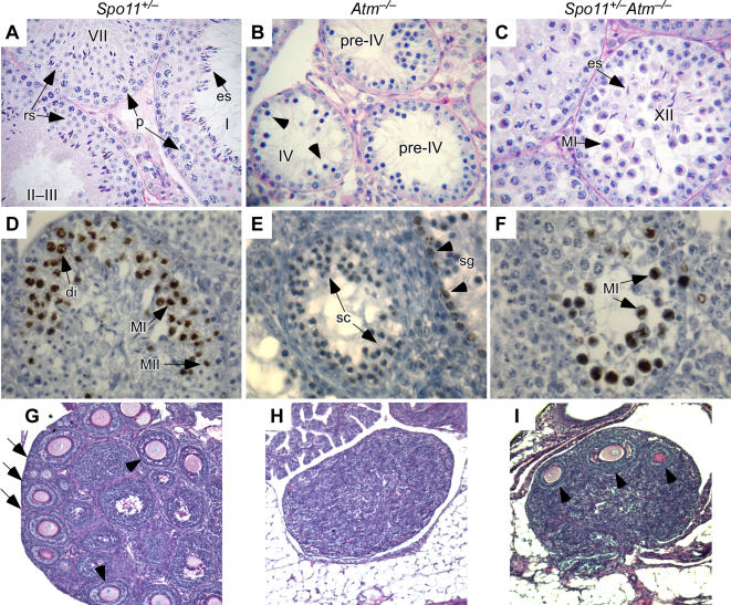
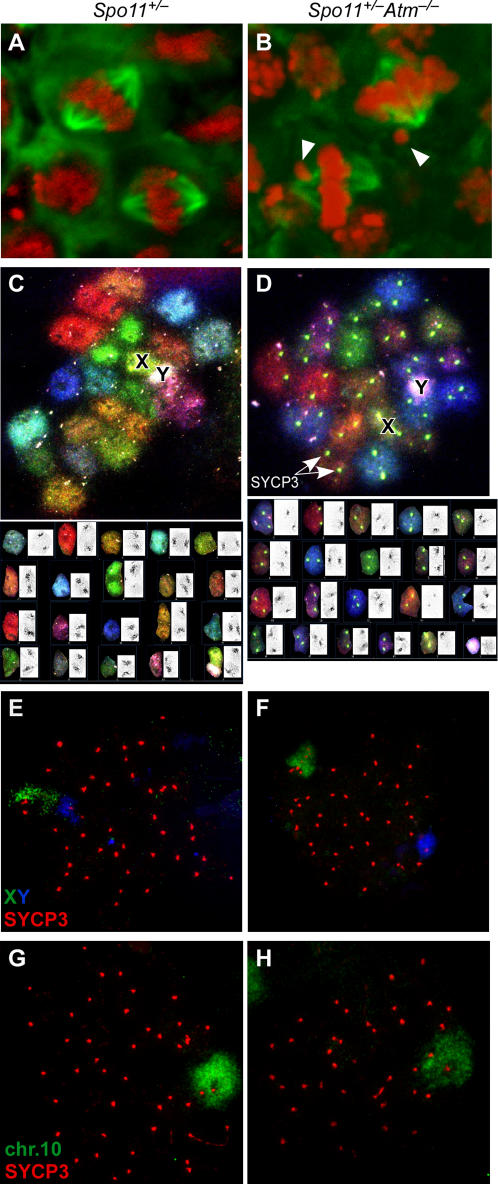
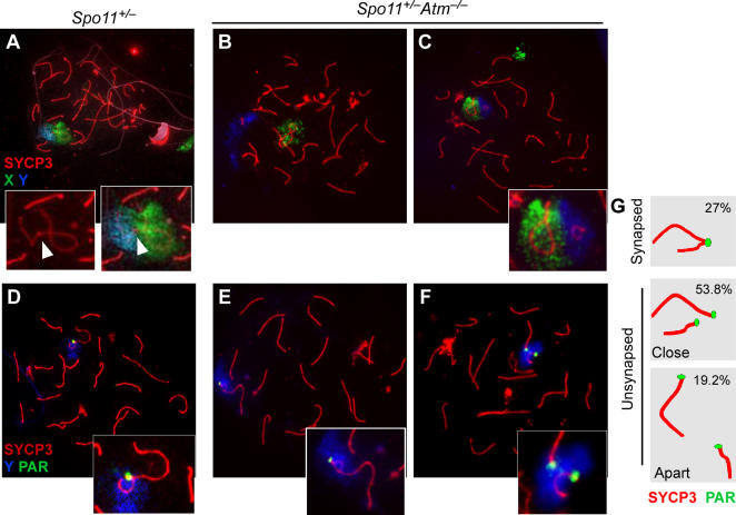
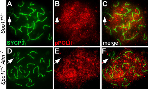
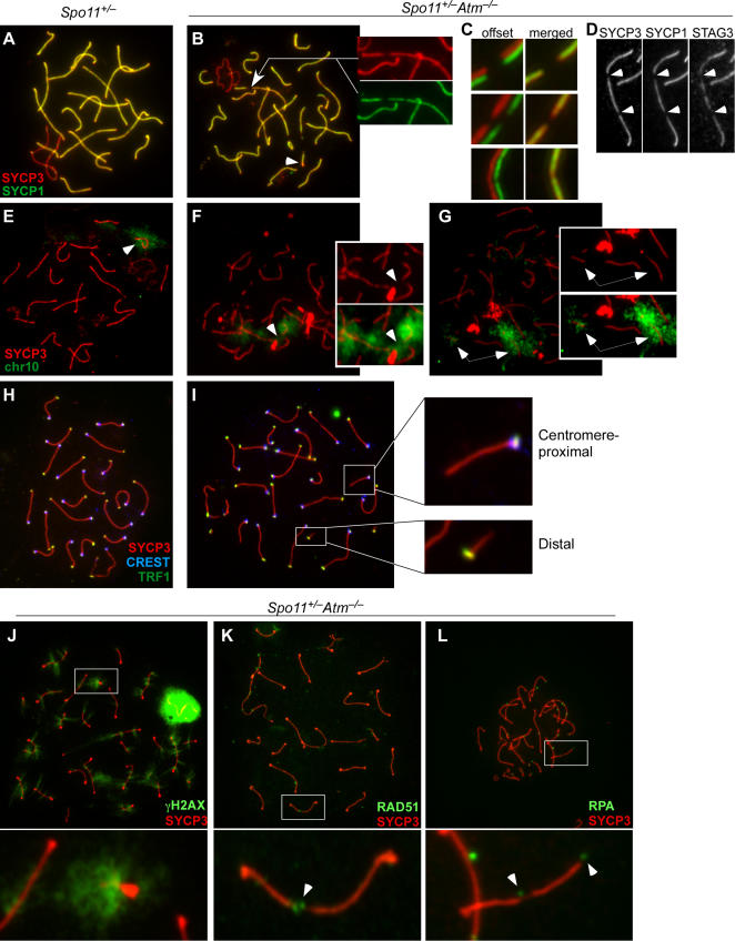
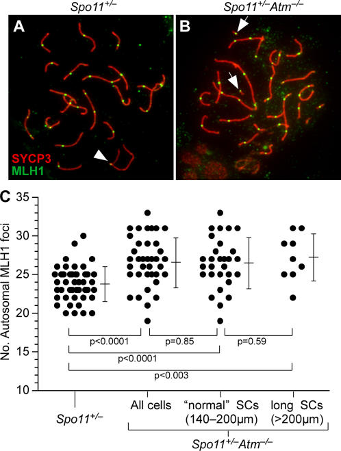
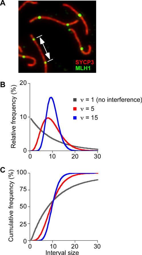
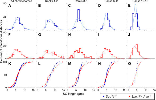
During meiosis in most sexually reproducing organisms, recombination forms crossovers between homologous maternal and paternal chromosomes and thereby promotes proper chromosome segregation at the first meiotic division. The number and distribution of crossovers are tightly controlled, but the factors that contribute to this control are poorly understood in most organisms, including mammals. Here we provide evidence that the ATM kinase or protein is essential for proper crossover formation in mouse spermatocytes. ATM deficiency causes multiple phenotypes in humans and mice, including gonadal atrophy. Mouse Atm-/- spermatocytes undergo apoptosis at mid-prophase of meiosis I, but Atm(-/-) meiotic phenotypes are partially rescued by Spo11 heterozygosity, such that ATM-deficient spermatocytes progress to meiotic metaphase I. Strikingly, Spo11+/-Atm-/- spermatocytes are defective in forming the obligate crossover on the sex chromosomes, even though the XY pair is usually incorporated in a sex body and is transcriptionally inactivated as in normal spermatocytes. The XY crossover defect correlates with the appearance of lagging chromosomes at metaphase I, which may trigger the extensive metaphase apoptosis that is observed in these cells. In addition, control of the number and distribution of crossovers on autosomes appears to be defective in the absence of ATM because there is an increase in the total number of MLH1 foci, which mark the sites of eventual crossover formation, and because interference between MLH1 foci is perturbed. The axes of autosomes exhibit structural defects that correlate with the positions of ongoing recombination. Together, these findings indicate that ATM plays a role in both crossover control and chromosome axis integrity and further suggests that ATM is important for coordinating these features of meiotic chromosome dynamics.


Conflict of interest statement

The authors have declared that no competing interests exist.

Figures

Figure 1
Figure 1. Spo11 heterozygosity partially ameliorates defects in meiotic progression in the absence of ATM.
(A–C) Periodic acid Schiff (PAS)-stained testis sections from mice of the indicated genotypes. Roman numerals denote stages of the seminiferous epithelial cycle . Spo11+/− mice (A) show normal patterns, with examples of pachytene spermatocytes (p), round spermatids (rs), and elongating spermatids (es) indicated. Atm−/− seminiferous tubules (B) show decreased cellularity as a result of spermatocyte apoptosis at stage IV. Cells that appear to be apoptotic are indicated (arrowheads). Spo11+/−Atm−/− tubules (C) have increased testis cellularity compared to Atm−/− mice. Examples are indicated of metaphase I spermatocytes (MI) and elongating spermatids (es) in stage XII tubules. The cytosol of many metaphase cells is intensely stained, suggesting that these cells are undergoing apoptosis (see also Figure S1). (D–F) Anti-phospho-histone H3 (p-H3) staining of testis sections. Presence of p-H3 is marked by a dark brown enzymatic precipitate. Examples of spermatogonia (sg) and spermatocytes in diplonema (di), metaphase I (MI), or metaphase II (MII) are indicated. Examples of the most advanced non-apoptotic spermatocytes observed in Atm−/− are indictated in panel E (sc). (G–I) Reduced Spo11 gene dosage partially rescues oogenesis in Atm−/− mice. Ovary sections from 17–18 dpp mice of the indicated genotypes were stained with PAS. In Spo11+/− mice (G), as in wild type, growing and antral follicles (arrowheads) and primordial follicles at the cortex of the ovary (arrows) are observed. Essentially no follicles at any stage of maturation are present in Atm−/− ovaries (H), whereas some growing and antral follicles (arrowheads) are observed in Spo11+/−Atm−/− ovaries (I).
Figure 2
Figure 2. ATM-deficient spermatocytes show metaphase I lagging chromosomes and defects in forming the obligate crossover between the sex chromosomes.
(A, B) Lagging chromosomes in Spo11+/−Atm−/− spermatocytes. Testis sections were immunostained with anti-tubulin antibodies to detect the spindle (green) and with DAPI (pseudocolored red). Arrowheads show examples of lagging chromosomes. (C, D) SKY of metaphase I chromosomes showing univalent X and Y in Spo11+/−Atm−/− (D) but not Spo11+/− (C) spermatocytes. Lower panels show karyotypes of the metaphases, with the inverted DAPI chromosome images in insets. Centromeres are visible as dark masses in the insets and as bright spots in (D), where anti-SYCP3 staining was included. (E–H) Metaphase I chromosome spreads from Spo11+/− (E, G) or Spo11+/−Atm−/− (F, H) were analyzed by FISH for the indicated chromosomes in conjunction with immunofluorescence for SYCP3. At metaphase I, SYCP3 is retained almost exclusively at the centromeric regions (although note that SYCP3 is also present as discrete foci along the X chromosome).
Figure 3
Figure 3. Aberrant sex chromosome synapsis in the absence of ATM.
Pachytene chromosome spreads from Spo11+/− (A, D) and Spo11+/−Atm−/− (B, C, E, F) were analyzed by immunofluorescence for SYCP3 along with FISH for X and Y (A–C) or Y and the PAR (D–F). Insets show the disposition of the X and Y. Arrowheads in (A) point to the synapsed PAR. (G) Frequencies of various sex chromosome configurations in Spo11+/−Atm−/− spreads based on FISH for Y and PAR. XY pairs were scored as “synapsed” if they showed a single PAR signal and intimately associated axes (e.g., panels D, E). If two PAR signals were observed, the X and Y were scored as “unsynapsed” and were further classified as to whether the chromosomes were close to one another in the spread (e.g., panel F) or far apart (similar to panel B).
Figure 4
Figure 4. ATM and XY chromosome synapsis are dispensable for meiotic sex chromosome inactivation.
Pachytene chromosome spreads were stained for SYCP3 and phosphorylated RNA polymerase II (pPOLII). In both Spo11+/− (A–C) and Spo11+/−Atm−/− (D–F), phosphorylated RNA polymerase II is excluded from the sex chromatin (arrows).
Figure 5
Figure 5. Chromosome structure defects in Spo11+/−Atm−/− spermatocytes.
(A–C) Axial element (SYCP3) and central element (SYCP1) defects in pachytene spermatocytes from Spo11+/–Atm–/– mice. Chromosome spreads from Spo11 +/– (A) and Spo11+/–Atm–/– spermatocytes (B–C) were immunostained with anti-SYCP3 and anti-SYCP1 antibodies. Insets in (B) highlight defects visible on some autosomes (arrow). Additional examples are shown in (C), which shows red and green immunofluorescence channels offset (left) or merged (right). (D) Colocalization of defects in SYCP3 and SYCP1 staining with gaps in staining for the meiotic cohesin subunit STAG3 in Spo11+/−Atm−/− spermatocytes. (E–G) Axial defects in Spo11+/−Atm−/− pachytene spermatocytes can be associated with chromosome fragmentation. Anti-SYCP3 immunofluorescence (red) and FISH for chr10 (green) was combined on spermatocyte spreads. In Spo11 +/− (E), SYCP3 and chr10 signals are always continuous. In Spo11+/−Atm−/−, examples are shown of SYCP3 gaps that either are not (F) or are (G) associated with overt fragmentation of chr10. (H, I) Short SC fragments are the distal ends of chromosomes. Spread pachytene nuclei from Spo11+/− (H) and Spo11+/−Atm−/− (I) were stained for SYCP3 (red) and the telomeric protein TRF1 (green), along with CREST antibodies to detect centromeres (blue). Insets in (I) show a short SC fragment from a distal chromosome end showing telomeric staining at one end (lower inset) and a longer SC fragment from a centromere-proximal chromosome end showing colocalization of the telomere and centromere (upper inset). (J–L) Colocalization of chromosome axis defects with markers of sites of DSB repair. Spo11+/−Atm−/− spermatocyte spreads were stained with anti-SYCP3 (red) and with antibodies to either γH2AX (J; 43 cells analyzed from 2 mice), RAD51 (K; 51 cells from 3 mice), or RPA (L; 42 cells from 2 mice). Insets show enlargements of the boxed regions of the spreads. Arrowheads point to examples of RAD51 or RPA foci that colocalize with SC gaps.
Figure 6
Figure 6. Elevated crossover numbers on autosomes in the absence of ATM.
(A, B) MLH1 foci on pachytene chromosomes. Spermatocyte spreads of the indicated genotype were stained for MLH1 (green) and SYCP3 (red). Arrowhead in (A) points to an MLH1 focus visible on the PAR; arrows in (B) show examples of MLH1 foci on short SC fragments. The XY pair at 6:00 o'clock in the spread in (B) does not show an MLH1 focus. (C) Autosomal MLH1 focus counts (bars = mean±sd). For Spo11+/−Atm−/−, data are shown for all of the cells analyzed (total cells, n = 38), the subset of cells (n = 29) that had a total SC length within the range observed in >90% of Spo11+/− cells (140–200 μm), and the subset of cells with longer SCs (>200 μm). P-values are shown for Mann-Whitney U tests of the indicated pairwise comparisons. The average total SC length (174.4±12.4 μm) in the subset of Spo11+/−Atm−/− cells with “normal” length SCs was slightly greater (3.5%) than the average SC length observed in Spo11+/− (167.7±16.3 μm), but this difference was not statistically significant (p = 0.065, t test).
Figure 7
Figure 7. Measuring cytological interference between MLH1 foci.
(A) Pachytene spermatocyte spreads were immunostained for SYCP3 (shown in red) and MLH1 (shown in green), then the distances between foci were measured on autosomal bivalents that contain two or more MLH1 foci. (B, C) Examples of relative (B) and corresponding cumulative (C) frequency plots of gamma distributions. If there is no interference between MLH1 foci, an exponential frequency distribution is expected (gray lines). Deviation from exponential behavior indicates the existence of interference (red and blue lines): short and long distances become more rare and the spacing becomes more even (i.e., distances are tightly clustered). Curves were calculated using an average interfocus distance of 10 and the indicated increasing values for the shape parameter, ν. See text and for further discussion.
Figure 8
Figure 8. Decreased cytological interference on autosomes in Spo11+/−Atm−/− spermatocytes.
Distances between pairs of adjacent MLH1 foci were measured on autosomes of pachytene spermatocytes. Panels A–E and F–J show the frequency distributions (step plots) of inter-focus distances for Spo11+/− (blue; 46 cells from 4 mice) and Spo11+/−Atm−/− (red; 38 cells from 4 mice), respectively. Best-fit gamma distributions are superimposed on each (smooth curves). Panels K–O show cumulative frequency plots to facilitate comparison of the two genotypes. The left column of graphs (A, F, K) pools data for all autosomes. The remaining columns show data for groups of similarly sized chromosomes, ranked from largest to smallest. Autosome size ranks 17–19 are excluded from this analysis because they rarely have more than a single MLH1 focus (see Table 1).

References

    1. Page SL, Hawley RS. Chromosome choreography: the meiotic ballet. Science. 2003;301:785–789. - PubMed
    1. Hillers KJ. Crossover interference. Curr Biol. 2004;14:R1036–R1037. - PubMed
    1. Jones GH, Franklin FC. Meiotic crossing-over: obligation and interference. Cell. 2006;126:246–248. - PubMed
    1. Perry J, Palmer S, Gabriel A, Ashworth A. A short pseudoautosomal region in laboratory mice. Genome Res. 2001;11:1826–1832. - PMC - PubMed
    1. Kleckner N, Zickler D, Jones GH, Dekker J, Padmore R, et al. A mechanical basis for chromosome function. Proc Natl Acad Sci U S A. 2004;101:12592–12597. - PMC - PubMed

Publication types

MeSH terms