. 2008 Jun;118(6):2025-38.
doi: 10.1172/JCI34267.

HIV protease inhibitors provide neuroprotection through inhibition of mitochondrial apoptosis in mice

Affiliations

HIV protease inhibitors provide neuroprotection through inhibition of mitochondrial apoptosis in mice

Toshio Hisatomi et al. J Clin Invest. 2008 Jun.

Abstract

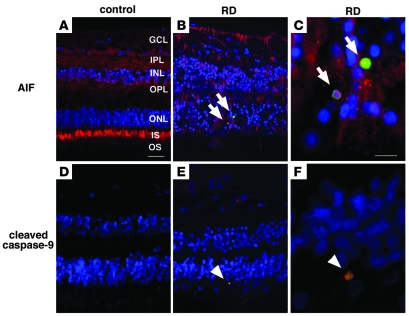
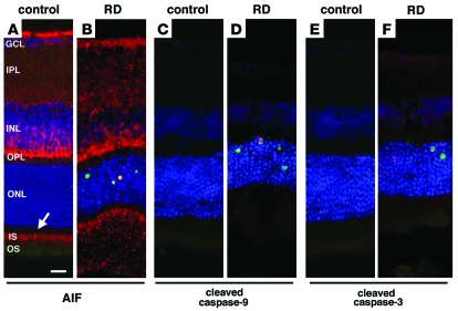
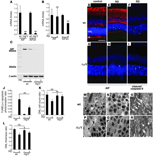
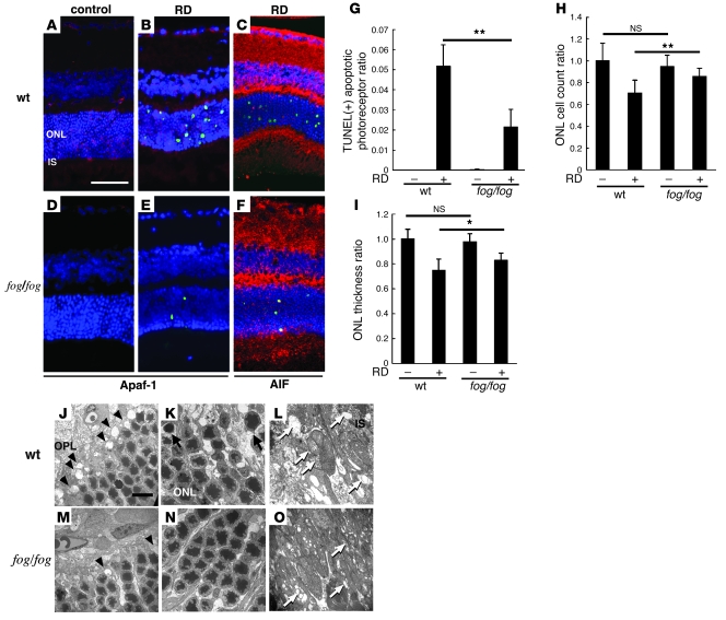
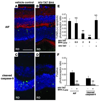
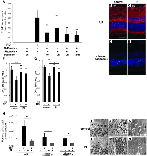
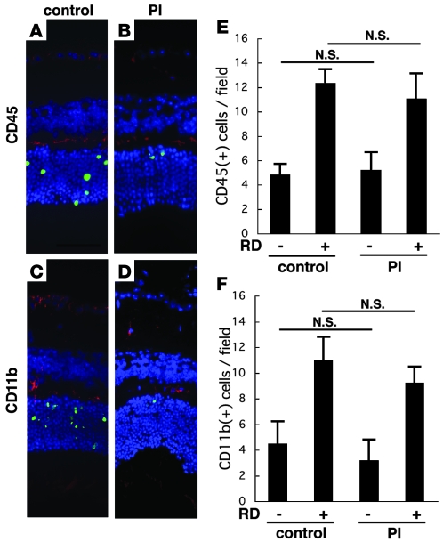
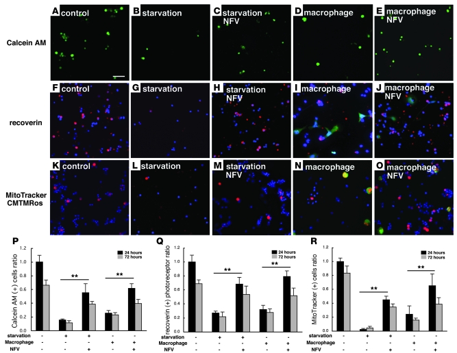
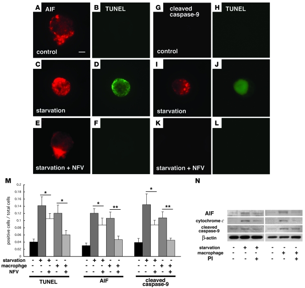
Neuroprotection can be achieved by preventing apoptotic death of postmitotic cells. Apoptotic death can occur by either a caspase-dependent mechanism, involving cytochrome c, apoptosis protease-activating factor-1 (Apaf-1), and caspase-9, or a caspase-independent mechanism, involving apoptosis-inducing factor (AIF). HIV protease inhibitors (PIs) avert apoptosis in part by preventing mitochondrial outer membrane permeabilization (MOMP), but the precise mechanism by which they work is not known. Here, we evaluated the impact of the PIs in a mouse model of retinal detachment (RD) in vivo and in murine primary retinal cell cultures in vitro. Oral administration of the PIs nelfinavir and ritonavir significantly inhibited photoreceptor apoptosis, while preventing the translocation of AIF from mitochondria to the nucleus as well as the activation of caspase-9. RD-induced photoreceptor apoptosis was similarly inhibited in mice carrying hypomorphic mutations of the genes encoding AIF or Apaf-1. Nelfinavir attenuated apoptosis as well as mitochondrial release of AIF and cytochrome c, and subsequent activation of caspase-9 in vitro, in photoreceptor cultures exposed to starvation or monocyte chemoattractant protein-1-stimulated (MCP-1-stimulated) macrophages. Our results suggest that the MOMP inhibition by PIs involved interruption of both caspase-dependent and caspase-independent apoptosis pathways and that PIs may be clinically useful for the treatment of diseases caused by excessive apoptosis.


Figures

Figure 1
Figure 1. Immunofluorescence detection of AIF and cleaved caspase-9 in the human retina after RD.
Paraffin-embedded retinal sections from the MEEI archives were analyzed retrospectively. Whereas histopathologic sections showed relatively preserved retinal structures in control retinas without RD (A and D), photoreceptor loss and secondary gliosis were observed in retinas with RD (B, C, E, and F). Specific AIF-staining was noted in ganglion cells (GCL), inner plexiform layer (IPL), inner nuclear layer (INL), outer plexiform layer (OPL), and photoreceptor inner segment (IS) and outer segment (OS) of control retina (A; AIF in red, TUNEL in green, DAPI in blue). Notably, in the detached retinas, AIF staining was mainly observed in proliferating glial cells as well as in TUNEL-positive photoreceptor nuclei (B and C, arrows). Although no staining was observed in control retinas without RD (D), cleaved caspase-9–positive cells were noted in the cytoplasm of TUNEL-positive cells in the detached retinas (cleaved caspase-9 in red, TUNEL in green, DAPI in blue; arrowheads in E and F). Scale bars: 50 μm (A, B, D, and E) and 10 μm (C and F).
Figure 2
Figure 2. Immunofluorescence detection of AIF and cleaved caspase-9 and -3 in the mouse retina after RD.
AIF-positive staining was noted in ganglion cells, inner plexiform layer, inner nuclear layer, outer plexiform layer, and photoreceptor inner segment (arrow) of control retinas (A; AIF in red, TUNEL in green, DAPI in blue). After RD, photoreceptors underwent apoptotic cell death, and AIF-positive staining was observed in TUNEL-positive photoreceptor nuclei (B). Cytosolic staining of cleaved caspase-9 (D, red) and -3 (F, red) was observed in the detached retinas but not in control attached retinas (C and E). Scale bar: 10 μm.
Figure 3
Figure 3. AIF deficiency protects from RD-induced photoreceptor apoptosis.
To examine the AIF expression in retinas of AIF mutant mice (Hq/Y), we compared the expression of AIF and AIFsh mRNA (A and B) and AIF protein (C) in WT and Hq/Y mice before and after RD. Hq/Y mice exhibited negligible levels of AIF mRNA or AIF protein in contrast to WT mice (AC). We also examined the expression of AIFsh, a proapoptotic short variant of AIF. The AIFsh mRNA expression was also decreased in AIF-deficient mice (Hq/Y; A and B), and AIFsh protein expression was not induced by RD in WT or Hq/Y mice (C). AIF immunohistochemistry confirmed the low AIF expression in Hq/Y mice (G and H; AIF in red, TUNEL in green, DAPI in blue) in contrast to WT mice (D and E). AIF deficiency in Hq/Y mice substantially decreased TUNEL-positive apoptotic cells after RD (E, H, and J) and preserved ONL cell count (K) and ONL thickness (L). The remaining TUNEL-positive apoptotic photoreceptors showed activated, cleaved caspase-9 in Hq/Y mice (F and I, caspase-9 in red). Electron microscopy also showed well-preserved rod spherules and cone pedicles in the outer plexiform layer (M and P, arrowheads; degenerated pedicles and spherules), decreased apoptotic nuclei in the ONL (N and Q, arrows), and relatively preserved mitochondria in the inner segment of photoreceptors (O and R, white arrows). n = 5 per group; *P < 0.05, **P < 0.01. Scale bars: 50 μm (D), 10 μm (M).
Figure 4
Figure 4. Apaf-1 deficiency protects from RD-induced photoreceptor apoptosis.
Apaf-1 expression was observed in WT mice (A and B; Apaf-1 in red, TUNEL in green, DAPI in blue) but deficient in Apaf-1 mutant fog/fog mice (D and E). TUNEL-positive apoptotic photoreceptor after RD decreased in fog/fog mice in contrast to WT mice (AG). The remaining TUNEL-positive apoptotic photoreceptors showed AIF translocation into the nucleus in fog/fog mice (C and F; AIF in red, TUNEL in green, DAPI in blue). In fog/fog mice, ONL cell count ratio (H) and ONL thickness ratio (I) were also preserved, in contrast to those in WT mice. Ultrastructural studies showed relatively well-preserved structures in the outer plexiform layer (J and M, arrowheads; rod spherules and cone pedicles), the ONL (K and N, arrows; apoptotic photoreceptors), and the inner segment of photoreceptors (L and O, white arrows; ruptured mitochondria). n = 5 per group; *P < 0.05, **P < 0.01. Scale bars: 50 μm (A), 10 μm (J).
Figure 5
Figure 5. Intraperitoneal injection of recombinant Bcl-XL fusion protein (HIV-TAT BH4) containing cell-permeable TAT protein transduction domain and antiapoptotic domain BH4 of Bcl-XL attenuated apoptotic photoreceptors after RD in a dose-dependent manner.
Notably, HIV-TAT BH4 protein also confined AIF translocation into the nucleus (A, B, and F; AIF in red, TUNEL in green, DAPI in blue) and decreased caspase-9 cleavage (C, D, and F; cleaved caspase-9 in red, TUNEL in green, DAPI in blue) after RD. Treatment of the animals with vehicle or a control cell-permeable HIV-TAT recombinant protein without the BH4 sequence did not affect photoreceptor apoptosis detected by TUNEL (E). n = 5 per group; **P < 0.01. Scale bar in A: 50 μm.
Figure 6
Figure 6. Systemic oral administration of HIV PIs prevents detachment-induced photoreceptor apoptosis as well as AIF translocation and caspase activation in vivo.
Each animal received vehicle (2% ethanol in dH20), single PI (NFV or RIT), or double PI (NFV boosted with RIT) 3 times daily, starting immediately after induction of RD (0h) or 24 hours after RD. Although NFV plus RIT did not affect control retina without RD, NFV and NFV/RIT substantially reduced TUNEL-positive apoptotic photoreceptors after RD (AE). NFV/RIT also blocked AIF translocation from mitochondria to nuclei (B, C, and H; AIF in red, TUNEL in green, DAPI in blue) and caspase-9 and -3 cleavage (D, E, and H; cleaved caspase-9 in red, TUNEL in green, DAPI in blue). PIs preserved the ONL cell count ratio (F) and ONL thickness ratio (G). NFV/RIT also maintained the ultrastructure of the retinas after RD (IN). In the outer plexiform layer (I and L), rod spherules and cone pedicles were well preserved in the NFV/RIT-treated group (L, arrowheads; degenerated pedicles and spherules) in contrast to the vehicle group (I). In the ONL, photoreceptors with characteristics of apoptosis (arrows), namely cellular shrinkage and chromatin condensation, decreased in the PI-treated group (M) in contrast to the vehicle-treated group (J). In the inner segment of photoreceptors, a large number of mitochondria was swollen and degenerated in the vehicle group (K, arrows); in contrast, mitochondria was well preserved in the PI-treated group (N). n = 5 per group; *P < 0.05, **P < 0.01. Scale bars: 50 μm (B), 10 μm (I).
Figure 7
Figure 7. Accumulation of CD45+ and CD11b+ myeloid lineage cells in the retina after RD.
To examine whether PIs might mediate their photoreceptor-protective effects indirectly, via suppression of microglial activity, we quantified the microglial infiltration/proliferation in the retina after RD by immunofluorescence detection of the leukocyte common antigen CD45 (A, B, and E; CD45 in red, TUNEL in green, DAPI in blue) and the macrophage/microglial marker CD11b (C, D, and F; CD11b in red, TUNEL in green, DAPI in blue) in control RD (A and C) and NFV/RIT-treated RD mice (B and D). Irrespective of the treatment with NFV/RIT, RD increased the frequency of CD45+ cells or CD11b+ cells in the outer plexiform layer (E and F). n = 5 per group.
Figure 8
Figure 8. PI increased survival of photoreceptors and preserved mitochondrial potential in vitro in 2 models of primary retinal cell cultures.
The viability of retinal cells was measured with calcein AM after starvation or after coculture with macrophages (AE and P; calcein AM in green). Calcein-positive viable cells decreased after starvation (B) or coculture (D) as compared with untreated control (A). PI (NFV, 7 μM) treatment significantly increased calcein-positive cells (C and E). Recoverin+ photoreceptors attaching on culture dish were analyzed after starvation or coculture with macrophages (FJ and Q; recoverin in red, GFP in green, DAPI in blue). Attaching recoverin+ photoreceptors decreased after 24 hours incubation (G and I) as compared with untreated control cultures (F). PI treatment significantly rescued photoreceptors from apoptotic cell death (H and J). Mitochondrial function was assessed by a mitochondrial potential–sensitive dye, CMTMRos. CMTMRos intake (KO, R; CMTMRos in red, GFP in green, DAPI in blue) was lost after induction of apoptosis (L and N) but reversed in NFV-treated cells (M and O). Representative images are shown (AO), and the quantified results are shown in PR. n = 10 per group; **P < 0.01. Scale bar: 100 μm.
Figure 9
Figure 9. PI blocked leakage of proapoptotic molecules from the mitochondria to the cytosol (AIF and cytochrome c) and subsequent activation of caspase-9 in vitro.
Representative images of immunofluorescence detection of AIF (AF; AIF in red, TUNEL in green) and cleaved activated caspase-9 (GL; cleaved caspase-9 in red, TUNEL in green) in control photoreceptor cultures (A, B, G, and H) and after starvation in the absence (C, D, I, and J) or presence of NFV (E, F, K, and L). Photoreceptors underwent TUNEL-detectable apoptosis in starvation and macrophage coculture models but were preserved by treatment with NFV (M). AIF translocation and caspase-9 cleavage were also decreased by NFV treatment (M). Immunoblot analyses of cytosolic fractions showed AIF and cytochrome c translocation from the mitochondria to the cytosol and subsequent caspase-9 cleavage under starvation or macrophage cocultures, and these changes were suppressed by NFV treatment (N). n = 10 per group; *P < 0.05, **P < 0.01. Scale bar in A: 1 μm.

References

    1. Dunaief J.L., Dentchev T., Ying G.S., Milam A.H. The role of apoptosis in age-related macular degeneration. Arch. Ophthalmol. 2002;120:1435–1442. - PubMed
    1. Barber A.J., et al. Neural apoptosis in the retina during experimental and human diabetes. Early onset and effect of insulin. J. Clin. Invest. 1998;102:783–791. doi: 10.1172/JCI2425. - DOI - PMC - PubMed
    1. Fulton A.B., Hansen R.M., Petersen R.A., Vanderveen D.K. The rod photoreceptors in retinopathy of prematurity: an electroretinographic study. Arch. Ophthalmol. 2001;119:499–505. - PubMed
    1. Cook B., Lewis G.P., Fisher S.K., Adler R. Apoptotic photoreceptor degeneration in experimental retinal detachment. Invest. Ophthalmol. Vis. Sci. 1995;36:990–996. - PubMed
    1. Berglin L., Algvere P.V., Seregard S. Photoreceptor decay over time and apoptosis in experimental retinal detachment. Graefes Arch. Clin. Exp. Ophthalmol. 1997;235:306–312. doi: 10.1007/BF01739640. - DOI - PubMed

Publication types