. 2008 Jun;237(6):1636-44.
doi: 10.1002/dvdy.21576.

Brd4 associates with mitotic chromosomes throughout early zebrafish embryogenesis

Affiliations

Brd4 associates with mitotic chromosomes throughout early zebrafish embryogenesis

Reiko Toyama et al. Dev Dyn. 2008 Jun.

Abstract

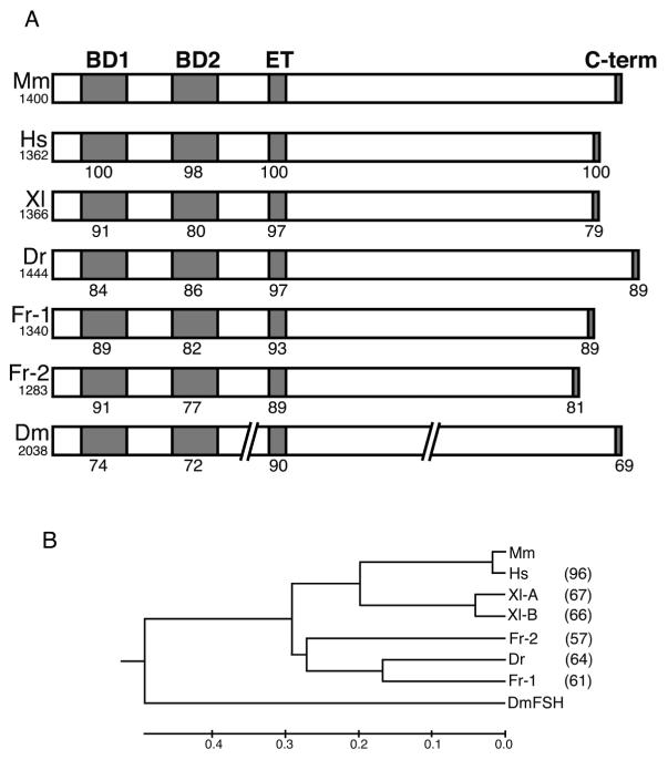
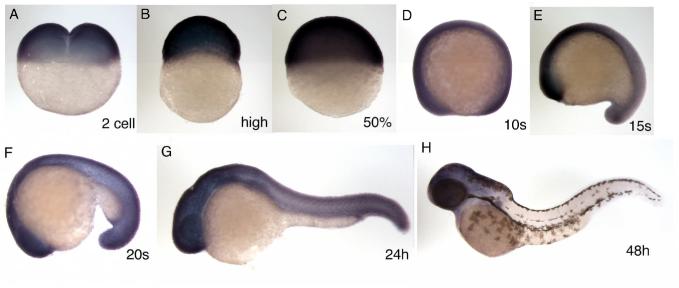
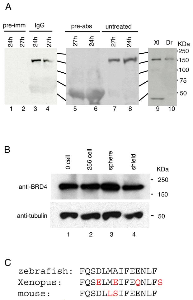
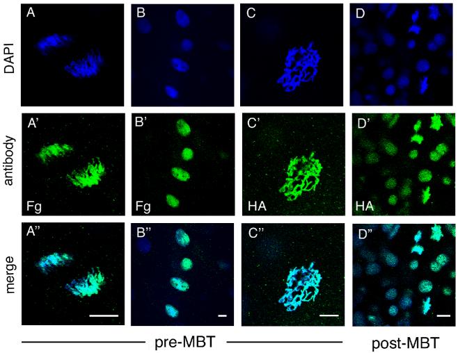
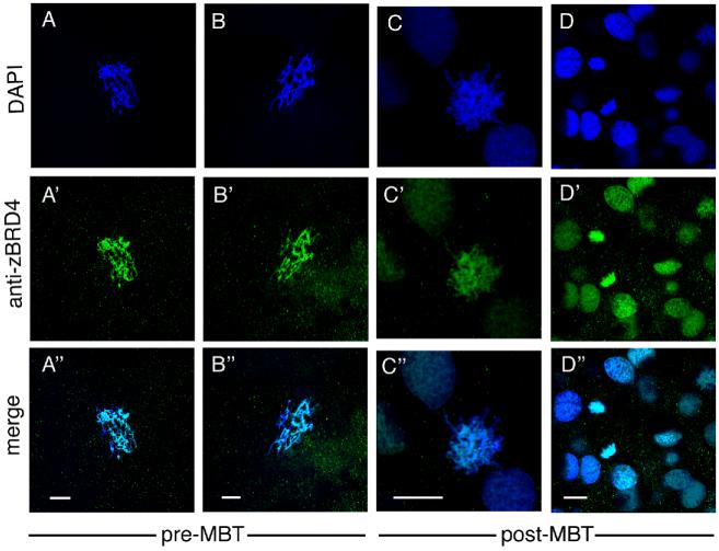
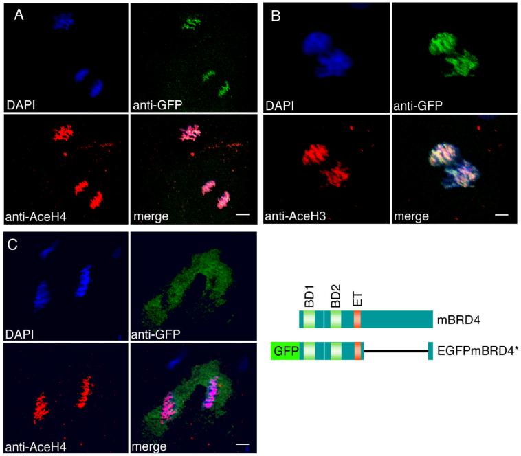
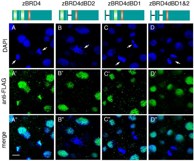
Brd4 is a member of the BET (bromodomains and extraterminal) subfamily of bromodomain proteins that includes chromatin-modifying proteins and transcriptional regulators. Brd4 has a role in cell cycle progression, making it indispensable in mouse embryos and cultured cells. The N-terminal domain of Brd4 participates in a fusion oncogene. Brd4 associates with acetylated histones in chromatin, and this association persists during mitosis implicating Brd4 in epigenetic memory. Brd4 sequence, particularly the bromodomains and ET domain, is conserved in the zebrafish and Xenopus laevis proteins reported here. Brd4 is expressed and localized on mitotic chromosomes in early zebrafish embryos before and after the midblastula transition (MBT), indicating that the Brd4-chromosome association is a conserved property that is maintained even before zygotic transcription. The association of Brd4 with acetylated histones may also be conserved in early embryos as we found that histones H3 and H4 are already acetylated during pre-MBT stages.


Figures

Fig. 1
Fig. 1
(A) Schematic structure of Brd4 proteins, containing two bromodomains, an ET domain, and a C-terminal domain. Amino acid identity with mouse Brd4 is indicated for each domain. Total amino acid length is shown under the name of the species. Mm, mouse; Hs, human; Xl, Xenopus; Dr, zebrafish; Fr, fugu; Dm, Drosophila. The Drosophila sequence is interrupted to indicate its substantially longer length. (B) Phylogenetic tree of Brd4 proteins. Numbers at the right indicate amino acid identity compared to the mouse. No value is entered for Drosophila because of the substantial length differences.
Fig. 2
Fig. 2
Expression pattern of the zebrafish brd4 gene. The stage examined by in situ hybridization, carried out as described by (Toyama and Dawid, 1997), is shown in each panel. A, 2 cell stage; B, high stage; C, 50% epiboly; D, 10 somite; E, 15 somite; F, 20 somite; G, 24 hpf; H, 48 hpf. All lateral view, anterior is left (D-H).
Fig. 3
Fig. 3
Brd4 protein analysis. (A) Antibody characterization. Polyclonal antibody against the Brd4 C-terminal 14 residues detected an approximately150 kD protein in zebrafish embryonic extracts (A, lanes 3, 4, 7, 8, and 10). Pre-immune serum (pre-imm, lanes 1 and 2) did not detect this protein. Antibody pre-incubated with antigen peptide (pre-abs, lane 5 and 6) failed to detect the 150 kD protein. Anti-Brd4 antibody cross-reacts with a protein of predicted size in extracts from Xenopus embryos (lane 9). (B) Brd4 protein during zebrafish embryogenesis. Extracts equivalent to two embryos were loaded in each lane. Lower panel shows α-tubulin as a loading control. Stages are indicated for each lane. (C) The C-terminal amino acid sequence of zebrafish, Xenopus, and mouse is shown with differences in red.
Fig. 4
Fig. 4
Localization of epitope-tagged Brd4 protein. RNA encoding HA- or FLAG-tagged zebrafish brd4 was injected into 1-2 cell stage embryos and visualized by antibody staining. Each column represents a separate embryo at the stage indicated. (A-A” and B-B”): FLAG-Brd4, (C-C” and D-D”): HA-Brd4. (A-C) Pre-MBT (512 cell stage) embryos with mitotic chromosomes (A, anaphase and C, prophase), and interphase nuclei (B) shown. (D) Post-MBT (sphere to dome stage) embryo presents both mitotic chromosomes and interphase nuclei in one embryo. Scale bar: 10 μm.
Fig. 5
Fig. 5
Localization of endogenous Brd4 protein visualized by staining with anti-zebrafish Brd4 antibody. (A-A” and B-B”): pre-MBT (256-512 cell stage) embryos, (C-C” and D-D”): post-MBT (sphere to dome stage) zebrafish embryos. A-C, prophase. Scale bar: 10 μm.
Fig. 6
Fig. 6
Brd4 protein is co-localized with acetylated histones in zebrafish embryos. (A, B) EGFP-mBrd4* RNA lacking aa 700-1317 was injected into 1-2 cell embryos; EGFP RNA was injected as control in (C). The embryos were fixed before MBT at 256 cell (A, C) and 128-256 cell stage (B), and immunostained with anti-acetyl-histone antibodies (red) and anti-GFP antibodies (green). (A) Anti-tetra-acetyl-histone H4; (B) anti-di-acetyl-histone H3; (C) anti-tetra-acetyl-histone H4. The structures of wild type and fusion brd4 constructs are shown. Scale bar: A and C, 8 μm; B, 4 μm.
Fig. 7
Fig. 7
Bromodomain-deleted zebrafish Brd4 proteins do not associate with mitotic chromosomes. Constructs are shown schematically at the top: Brd4, FLAG-tagged construct containing both bromodomains; constructs with deletions of bromodomain 1 (dBD1), bromodomain 2 (dBD2), or both bromodomains (dBD1 and 2). RNAs were injected into 1-2 cell stage zebrafish embryos, and stained at the sphere stage with anti-FLAG antibody (green). Arrows indicate mitotic chromosomes. Scale bar: μm.

References

    1. Aparicio S. Vertebrate evolution: recent perspectives from fish. Trends Genet. 2000;16:54–6. - PubMed
    1. Bisgrove DA, Mahmoudi T, Henklein P, Verdin E. Conserved P-TEFb-interacting domain of BRD4 inhibits HIV transcription. Proc Natl Acad Sci U S A. 2007;104:13690–5. - PMC - PubMed
    1. Chang YL, King B, Lin SC, Kennison JA, Huang DH. A double-bromodomain protein, FSH-S, activates the homeotic gene ultrabithorax through a critical promoter-proximal region. Mol Cell Biol. 2007;27:5486–98. - PMC - PubMed
    1. Chua P, Roeder GS. Bdf1, a yeast chromosomal protein required for sporulation. Mol Cell Biol. 1995;15:3685–96. - PMC - PubMed
    1. Clute P, Masui Y. Regulation of the appearance of division asynchrony and microtubule-dependent chromosome cycles in Xenopus laevis embryos. Dev Biol. 1995;171:273–85. - PubMed

Publication types