

doi: 10.1002/prot.22095.
Crystal structure of a putative lysostaphin peptidase from Vibrio cholerae
Affiliations
- PMID: 18498110
- PMCID: PMC3614409
- DOI: 10.1002/prot.22095
Item in Clipboard
Crystal structure of a putative lysostaphin peptidase from Vibrio cholerae
Proteins.
.
No abstract available
Figures
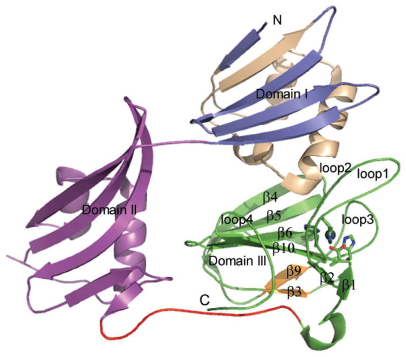
Ribbons representation of putative lysostaphin peptidase from Vibrio cholerae. The three structural domains are labeled. N-terminal residues (11–57) of Domain I resembling LysM structural motif are shown in wheat color. In Domain III, strands β3 and β9 which are insertions to lysostaphin type peptidase are shown in orange. The active site residues and the zinc ion are shown as stick and ball model, respectively. The long loop connecting the N and C domains is shown in red.

Stereoview of C-terminal domains of P01, P02 and active LytM superimposed. P01, P02, and LytM are shown in green, purple and cyan, respectively. Loops 2 and 4 of LytM move toward the central β-core region compared to P01 and P02.
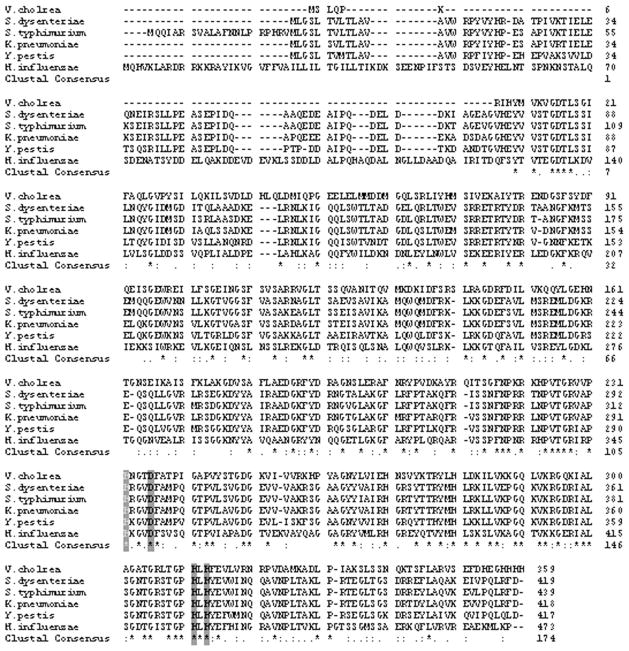
Sequence alignment of putative peptidase from Vibrio cholerae (P01) homologs in human pathogenic bacteria like Shigella dysenteriae (GI:82776442), Klebsiella pneumoniae (GI:152970918), Salmonella typhimurium (GI:16765232), Yersinia pestis (GI:16122301), Haemophilus influenzae (GI:145636322). The conserved residues are shown in “*”and the similar sequences are shown “.” The four conserved active site residues are highlighted in gray. N-terminal region of V. cholerae matches with other human pathogenic bacteria.

A cartoon representation of N-terminal long α-helix (blue) and the surface representation of the C-terminal catalytic domain with the ribbons representation in laid (brown). The gray sphere represents Zn ion.
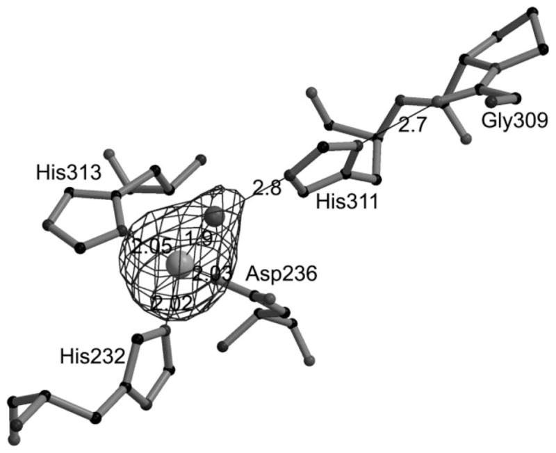
C-terminal domain active site residues are shown in ball and stick model. Contour represents Sigma weighted Fo-Fc map at 3.0 σ for Zn and catalytic water molecule. The dotted lines represent the interaction between the atoms and their distances are in Å unit.
References
-
- Hoffmann JA. The immune response of Drosophila. Nature. 2003;426:33–38. - PubMed
-
- Medzhitov R, Janeway CA., Jr Decoding the patterns of self and nonself by the innate immune system. Science. 2002;296:298–300. - PubMed
-
- Doyle RJ, Dziarski R. The bacterial cell: peptidoglycan. In: Sussman M, editor. Molecular medical microbiology. 7. Vol. 1. 2001. pp. 137–153.
-
- van Heijenoort J. Formation of the glycan chains in the synthesis of bacterial peptidoglycan. Glycobiology. 2001;11:25R–36R. - PubMed
-
- Stewart-Tull DE. Major component of the cell wall in gram positive organisms. Consists of a glycan backbone with alternating b 1–4 linked residues of N-acetyl-D-glucosamine and muramic acid. The immunological activities of bacterial peptidoglycans. Ann Rev Microbiol. 1980;34:311. - PubMed
Publication types
MeSH terms
Substances
Associated data
- Actions
- Actions
- Actions
Grants and funding
LinkOut - more resources
Full Text Sources
