

Cell encapsulation in biodegradable hydrogels for tissue engineering applications
- PMID: 18498217
- PMCID: PMC2962861
- DOI: 10.1089/ten.teb.2007.0332
Cell encapsulation in biodegradable hydrogels for tissue engineering applications
Abstract
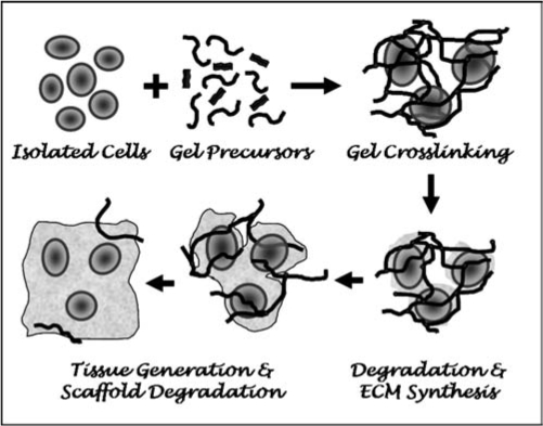
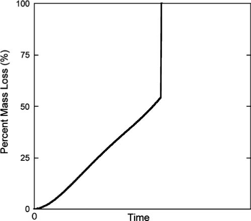
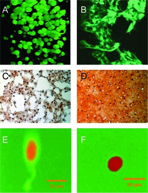
Encapsulating cells in biodegradable hydrogels offers numerous attractive features for tissue engineering, including ease of handling, a highly hydrated tissue-like environment for cell and tissue growth, and the ability to form in vivo. Many properties important to the design of a hydrogel scaffold, such as swelling, mechanical properties, degradation, and diffusion, are closely linked to the crosslinked structure of the hydrogel, which is controlled through a variety of different processing conditions. Degradation may be tuned by incorporating hydrolytically or enzymatically labile segments into the hydrogel or by using natural biopolymers that are susceptible to enzymatic degradation. Because cells are present during the gelation process, the number of suitable chemistries and formulations are limited. In this review, we describe important considerations for designing biodegradable hydrogels for cell encapsulation and highlight recent advances in material design and their applications in tissue engineering.
Figures
References
-
- Peretti G.M. Campo-Ruiz V. Gonzalez S. Randolph M.A. Xu J.W. Morse K.R. Roses R.E. Yaremchuk M.J. Tissue engineered cartilage integration to live and devitalized cartilage: a study by reflectance mode confocal microscopy and standard histology. Connect. Tissue Res. 2006;47:190. - PubMed
-
- Peppas N.A. Hilt J.Z. Khademhosseini A. Langer R. Hydrogels in biology and medicine: from molecular principles to bionanotechnology. Adv. Mater. 2006;18:1345.
-
- Mikos A.G. Papadaki M.G. Kouvroukoglou S. Ishaug S.L. Thomson R.C. Islet transplantation to create a bioartificial pancreas—mini review. Biotechnol. Bioeng. 1994;43:673. - PubMed
-
- Cao Y.L. Rodriguez A. Vacanti M. Ibarra C. Arevalo C. Vacanti C.A. Comparative study of the use of poly(glycolic acid), calcium alginate and pluronics in the engineering of autologous porcine cartilage. J. Biomater. Sci. Polym. Ed. 1998;9:475. - PubMed
-
- Sims C.D. Butler P.E. Cao Y.L. Casanova R. Randolph M.A. Black A. Vacanti C.A. Yaremchuk M.J. Tissue engineered neocartilage using plasma derived polymer substrates and chondrocytes. Plast. Reconstr. Surg. 1998;101:1580. - PubMed
Publication types
MeSH terms
Substances
Grants and funding
LinkOut - more resources
Full Text Sources
Other Literature Sources
