. 2008 Jul 1;198(1):143-9.
doi: 10.1086/588819.

Detection of epithelial-cell injury, and quantification of infection, in the HCT-8 organoid model of cryptosporidiosis

Affiliations

Detection of epithelial-cell injury, and quantification of infection, in the HCT-8 organoid model of cryptosporidiosis

Cirle Alcantara Warren et al. J Infect Dis. .

Abstract

Background: Intestinal cells grown in microgravity produce a three-dimensional tissue assembly, or "organoid," similar to the human intestinal mucosa, making it an ideal model for enteric infections such as cryptosporidiosis.

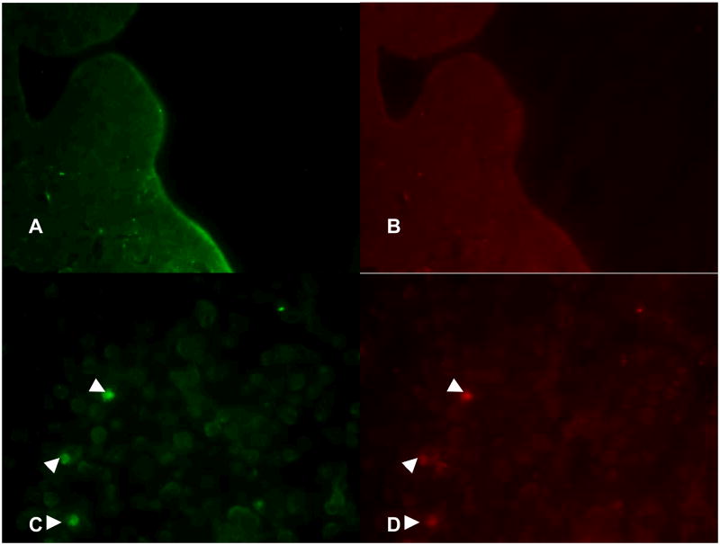
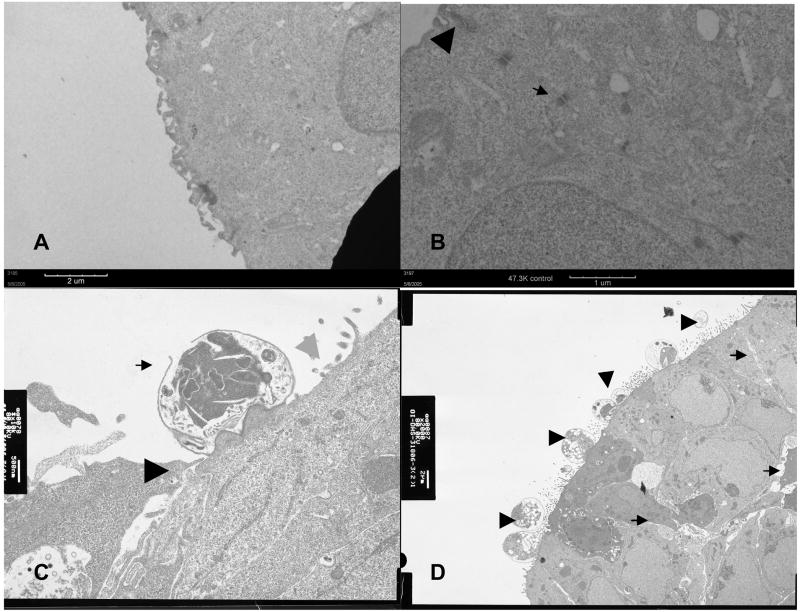
Methods: HCT-8 cells were grown in a reduced-gravity, low-shear, rotating-wall vessel (RWV) and were infected with Cryptosporidium parvum oocysts. Routine and electron microscopy (EM), immunolabeling with fluorescein-labeled Vicia villosa lectin and phycoerythrin-labeled monoclonal antibody to a 15-kD surface-membrane protein, and quantitative polymerase chain reaction (qPCR) using probes for 18s rRNA of C. parvum and HCT-8 cells were performed.

Results: The RWV allowed development of columnar epithelium-like structures. Higher magnification revealed well-developed brush borders at the apical side of the tissue. Incubation with C. parvum resulted in patchy disruption of the epithelium and, at the surface of several epithelial cells, in localized infection with the organism. EM revealed irregular stunting of microvilli, foci of indistinct tight junctions, and areas of loose paracellular spaces. qPCR showed a 1.85-log (i.e., 70-fold) progression of infection from 6 h to 48 h of incubation.

Conclusion: The HCT-8 organoid displayed morphologic changes indicative of successful and quantifiable infection with C. parvum. The HCT-8 organoid-culture system may have application in interventional in vitro studies of cryptosporidiosis.


Conflict of interest statement

All authors do not have any commercial or other association that might pose a conflict of interest.

Figures

Figure 1
Figure 1
Low power microscopy of HCT-8 cells organoid showing villus-like epithelial folds around the bioscaffold (A). At high power magnification, the HCT-8 cells formed layers of cells with the outermost layer showing polarization of cells and development of apical brush borders. Underneath the HCT-8 cell layers were loosely organized undifferentiated HCT-8 cells (B). Infected organoid showing localized infection and detachment of both uninfected and infected cells in the affected area (C). Oil power magnification showed disruption of apical brush borders (arrows) and looseness of intercellular attachments (arrowheads) (D). There were several detached cells, including possibly apoptotic cells, within the vicinity.
Figure 2
Figure 2
Fluorescent imaging of infected and uninfected HCT-8 cells. Panels A and B show uninfected cells stained with fluorescein-labeled Vicia villosa lectin specific to intracellular C. parvum sporozoites and phycoerythrin-labeled monoclonal antibody against the 15 kD membrane protein of C. parvum, respectively. Panels C and D showed intracellular C. parvum-positive cells (arrowheads) in the infected organoids.
Figure 3
Figure 3
Electron micrographs of uninfected organoids showed well-developed microvilli (Panel A) and distinct junctional apparatuses, including tight junction (arrowhead) and desmosomes (arrow) (Panel B). Panel C shows infected organoids with a meront (arrow), probably in the process of releasing merozoites, indistinct tight junction (black arrowhead) and denudation of the apical surface (gray arrowhead). Panel D shows an area of multiple infection with C. parvum at different stages of development (arrowheads) and loosening of the paracellular spaces (arrows). Of note, the tight junctions and desmosomes became inapparent in the infected organoids.
Figure 4
Figure 4
Comparison of infection rates between HCT-8 organoids and monolayers using quantitative PCR with C. parvum and HCT-8 cell probes. The organoids showed a steady increase in infection from 6 to 48 hours. The infection rates were higher in monolayers (Panel B) than in organoids (Panel A) but the rate of increase was more pronounced in the organoids than in the monolayers. On extended incubation, after the peak of infection at 48 hours, a steady decrease in parasite burden is noted starting at 76 hours post-infection (Panel C). Decrease in HCT-8 cells occurred at 96 hours suggesting cell detachment from the bioscaffold in infected organoids compared with the more stable amount of cells in the intact, uninfected organoids.

References

    1. Adams RB, Guerrant RL, Zu S, Fang G, Roche JK. Cryptosporidium parvum infection of intestinal epithelium: morphologic and functional studies in an in vitro model. J Infect Dis. 1994 Jan;169(1):170–7. - PubMed
    1. Griffiths JK, Moore R, Dooley S, Keusch GT, Tzipori S. Cryptosporidium parvum infection of Caco-2 cell monolayers induces an apical monolayer defect, selectively increases transmonolayer permeability, and causes epithelial cell death. Infect Immun. 1994 Oct;62(10):4506–14. - PMC - PubMed
    1. Upton SJ, Tilley M, Brillhart DB. Comparative development of Cryptosporidium parvum (Apicomplexa) in 11 continuous host cell lines. FEMS Microbiol Lett. 1994 May 15;118(3):233–6. - PubMed
    1. Laurent F, McCole D, Eckmann L, Kagnoff MF. Pathogenesis of Cryptosporidium parvum infection. Microbes Infect. 1999 Feb;1(2):141–8. - PubMed
    1. Seydel KB, Zhang T, Champion GA, et al. Cryptosporidium parvum infection of human intestinal xenografts in SCID mice induces production of human tumor necrosis factor alpha and interleukin-8. Infect Immun. 1998 May;66(5):2379–82. - PMC - PubMed

Publication types