. 2008 Oct;18(4):474-83.
doi: 10.1111/j.1750-3639.2008.00135.x. Epub 2008 May 22.

Hypocretin and melanin-concentrating hormone in patients with Huntington disease

Affiliations

Hypocretin and melanin-concentrating hormone in patients with Huntington disease

Ahmad Aziz et al. Brain Pathol. 2008 Oct.

Abstract

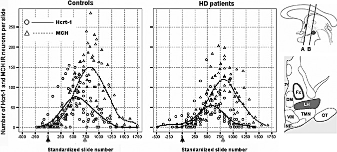
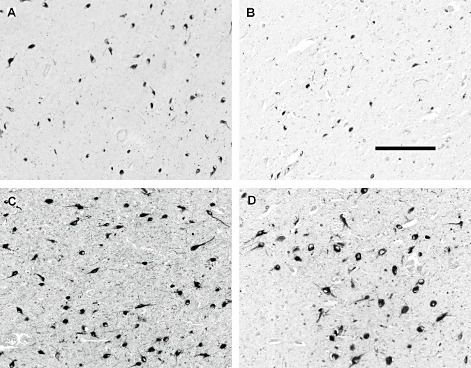
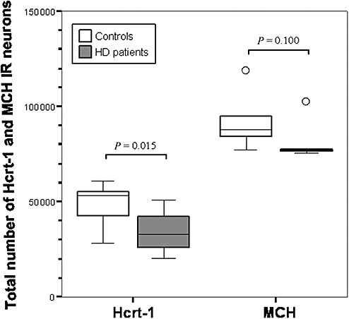
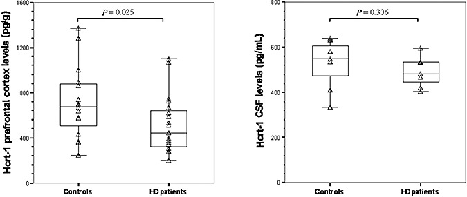
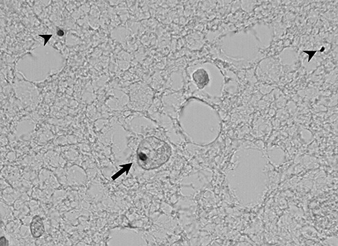
To evaluate whether hypocretin-1 (orexin-A) and melanin-concentrating hormone (MCH) neurotransmission are affected in patients with Huntington disease (HD), we immunohistochemically stained hypocretin and MCH neurons and estimated their total numbers in the lateral hypothalamus of both HD patients and matched controls. In addition, hypocretin-1 levels were determined in prefrontal cortical tissue and post-mortem ventricular cerebrospinal fluid (CSF) using a radioimmunoassay. The total number of hypocretin-1 neurons was significantly reduced by 30% in HD brains (P = 0.015), while the total number of MCH neurons was not significantly altered (P = 0.100). Levels of hypocretin-1 were 33% lower in the prefrontal cortex of the HD patients (P = 0.025), but ventricular CSF levels were similar to the control values (P = 0.306). Neuronal intranuclear and cytoplasmic inclusions of mutant huntingtin were present in all HD hypothalami, although with a variable distribution across different hypothalamic structures. We found a specific reduction in hypocretin signaling in patients with HD as MCH cell number was not significantly affected. It remains to be shown whether the moderate decrease in hypocretin neurotransmission could contribute to clinical symptoms. As the number of MCH-expressing neurons was not affected, alterations in MCH signaling are unlikely to have clinical effects in HD patients.


Figures

Figure 1
Figure 1
Distribution patterns of hypocretin‐1 (Hcrt‐1) and melanin‐concentrating hormone (MCH) immunoreactive (IR) neurons in the lateral hypothalamus of the controls and the Huntington disease (HD) patients. In order to present data from all subjects in the same diagrams, the individual distribution patterns were standardized in rostrocaudal direction for the anatomic distance between the point where the fornix abuts the paraventricular nucleus (black arrows; at the level of line “A” in the upper cartoon) and the fornicomamillary junction (grey arrows; at the level of line “B” in the upper cartoon). This procedure was performed separately for the control and the HD group. In addition, local linear regression was used to fit curves to the standardized pooled data in order to clarify the underlying distribution patterns (kernel = Gaussian, bandwidth value = 1.0). Note that the overall rostrocaudal dispersion of hypocretin‐1 and MCH IR neurons does not appear to be noticeably different between the control and the HD subjects. Upper cartoon. A schematic sagittal view of the hypothalamus; the paraventricular nucleus and the mamillary bodies are indicated in dark. Lower cartoon. A schematic coronal view of the hypothalamus midway between lines “A” and “B” in the upper cartoon; the perifornical area and the lateral hypothalamus are indicated in dark [adapted from (36)]. 3V = third ventricle; Fx = fornix; LH = lateral hypothalamus; DM = dorsomedial nucleus; VM = ventromedial nucleus; TMN = tuberomamillary nucleus; INF = infundibular nucleus; OT = optic tract.
Figure 2
Figure 2
Representative photographs of hypocretin‐1 and melanin‐concentrating hormone (MCH) immunoreactive (IR) neurons in the lateral hypothalamus of two control subjects (A and C, #C‐4 and #C‐2 respectively) and two patients with Huntington disease (HD) (B and D, #HD‐6 and #HD‐1 respectively) with neuronal counts at or around the median (Table 1). The pictures were taken from the slides with the highest numbers of IR neurons. The illustrations show a modest reduction in the number of hypocretin‐1 IR neurons in the HD brain (A,B) while the number of MCH IR neurons is not significantly affected (C,D). Scale bar: 250 µm.
Figure 3
Figure 3
The total numbers of hypocretin‐1 (Hcrt‐1) and melanin‐concentrating hormone (MCH) immunoreactive (IR) neurons in the lateral hypothalamus of control subjects and Huntington disease (HD) patients. The total number of Hcrt‐1 IR neurons is significantly decreased in HD, while there is only a trend toward a reduction in the total number of MCH IR neurons (Mann–Whitney U‐test: n = 16, P = 0.015 for Hcrt‐1 and n = 11, P = 0.100 for MCH). Outliers (defined as data points which lie 1.5 times the interquartile range below the first or above the third quartile) are symbolized by “○.”
Figure 4
Figure 4
Compared with the controls, hypocretin‐1 (Hcrt‐1) levels were significantly lower in the prefrontal cortex of the Huntington disease (HD) patients (Mann–Whitney U‐test (MWU): P = 0.025, left panel), but not in their cerebrospinal fluid (CSF) (MWU‐test: P = 0.306, right panel).
Figure 5
Figure 5
Examples of neuronal intranuclear (arrow) and cytoplasmic (arrowheads) inclusions of mutant huntingtin in the tuberomamillary nucleus of one Huntington disease patient (#HD‐7; obj. ×60 oil).

References

    1. Amiot C, Brischoux F, Colard C, La Roche A, Fellmann D, Risold PY (2005) Hypocretin/orexin‐containing neurons are produced in one sharp peak in the developing ventral diencephalon. Eur J Neurosci 22:531–534. - PubMed
    1. Aziz NA, Swaab DF, Pijl H, Roos RAC (2007) Hypothalamic dysfunction and neuroendocrine and metabolic alterations in Huntington disease: clinical consequences and therapeutic implications. Rev Neurosci 18:223–252. - PubMed
    1. Baumann CR, Hersberger M, Bassetti CL (2006) Hypocretin‐1 (orexin A) levels are normal in Huntington's disease. J Neurol 253:1232–1233. - PubMed
    1. Björkqvist M, Petersen A, Nielsen J, Ecker D, Mulder H, Hayden M et al (2006) Cerebrospinal fluid levels of orexin‐A are not a clinically useful biomarker for Huntington disease. Clin Genet 70:78–79. - PubMed
    1. Brundin L, Petersen A, Björkqvist M, Traskman‐Bendz L (2007) Orexin and psychiatric symptoms in suicide attempters. J Affect Disord 100:259–263. - PubMed

Publication types

MeSH terms