Review
. 2008 May 22;58(4):472-97.
doi: 10.1016/j.neuron.2008.04.030.

Glutamate receptor dynamics in dendritic microdomains

Affiliations
Review

Glutamate receptor dynamics in dendritic microdomains

Thomas M Newpher et al. Neuron. .

Abstract

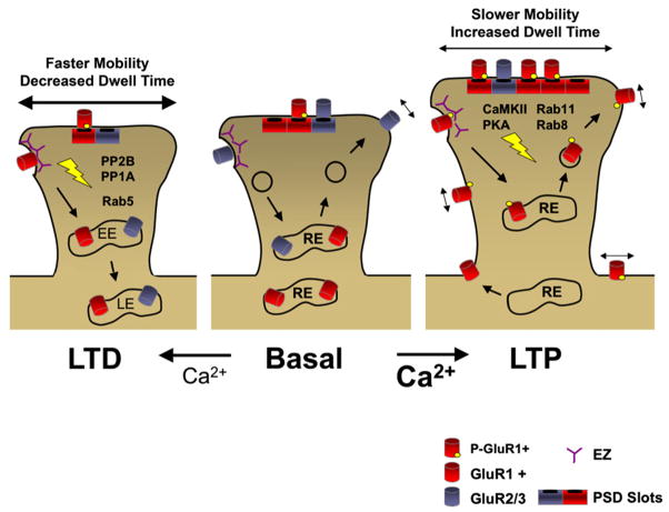
Among diverse factors regulating excitatory synaptic transmission, the abundance of postsynaptic glutamate receptors figures prominently in molecular memory and learning-related synaptic plasticity. To allow for both long-term maintenance of synaptic transmission and acute changes in synaptic strength, the relative rates of glutamate receptor insertion and removal must be tightly regulated. Interactions with scaffolding proteins control the targeting and signaling properties of glutamate receptors within the postsynaptic membrane. In addition, extrasynaptic receptor populations control the equilibrium of receptor exchange at synapses and activate distinct signaling pathways involved in plasticity. Here, we review recent findings that have shaped our current understanding of receptor mobility between synaptic and extrasynaptic compartments at glutamatergic synapses, focusing on AMPA and NMDA receptors. We also examine the cooperative relationship between intracellular trafficking and surface diffusion of glutamate receptors that underlies the expression of learning-related synaptic plasticity.


Figures

Figure 1
Figure 1. Nanoarchitecture of the PSD and Single-Particle Tracking of AMPA Receptors in Synaptic and Extrasynaptic Compartments
(A) AMPA receptor trajectories within synaptic and extrasynaptic compartments. As a control, immobilized Cy5-anti-GluR2 was fixed onto a coverslip (1). Examples 2–5 are trajectories from tracking of single Cy5-anti-GluR2 bound to AMPA receptors on living dendrites. The trajectories recorded in synaptic and extrasynaptic regions are shown in green and red, respectively. Examples 2 and 3 remained within synaptic sites, example 4 remained in the extrasynaptic membrane, and example 5 began in the extrasynaptic region and entered into a synaptic site. (B) Plots of the mean square displacement (MSD) versus time corresponding to the examples shown in (A). Trajectories 2 and 3 remaining in synaptic regions had varying degrees of confinement and were less mobile than trajectory 4. Error bars are equal to the SEM. (A) and (B) are adapted from Tardin et al. (2003); reprinted with permission from Nature Publishing Group, copyright 2003. (C) Individual GluR1-QDs are restricted to subdomains within active synapses. Five individual synaptic regions defined as a set of connected pixels are indicated. Individual pixels divided into 0.0016 μm2 subdomains were coded based on the presence (pink) or absence (white) of a GluR1-QD residing in that location at any time during the imaging period as defined by the centroid of a 2D Gaussian function fit to the GluR1-QD fluorescent signal. Scale bar, 200 nm. Adapted from Ehlers et al. (2007); reprinted with permission from Elsevier, copyright 2007. (D) CaMKII immunogold labeling (white dots) on the cytoplasmic surface of a biochemically isolated PSD. Shown is the cytoplasmic surface of the PSD. A membrane patch is indicated by the arrowhead. Scale bar, 100 nm. Adapted from Petersen et al. (2003); reprinted with permission from the Society for Neuroscience, copyright 2003. (E) AMPA receptor distribution at synapses (colored in red) in the molecular layer of the cerebellum shown by SDS-digested freeze-fracture replica labeling. Intra-membrane particles are shown on the E face of the PSD and contain dark immunogold particles for pan-AMPA receptors (GluR1-4). Scale bar, 100 nm. Adapted from Masugi-Tokita et al. (2007); reprinted with permission from the Society for Neuroscience, copyright 2007.
Figure 2
Figure 2. Glutamate Receptor Interactions in Synaptic, Perisynaptic, and Extrasynaptic Compartments
Distinct protein networks engage AMPA receptors and NMDA receptors in synaptic, perisynaptic, and extrasynaptic compartments. Direct protein interactions are shown by connecting lines. See text for details.
Figure 3
Figure 3. Active Synapses Capture GluR1-QDs by Diffusional Exchange
(A) Diffusion of GluR1 from silenced to active synapses. Trajectories of GluR1-QDs originating in a silenced synapse (green), escaping from the synapse (black, extrasynaptic), and transiting to an active synapse (red). The trajectory starts at point (a) and ends at point (b). (B) Plots of the instantaneous diffusion coefficient versus time for the examples in (A). Bars correspond to extrasynaptic (black), silenced synapses (green), and active synapses (red). (C) A model for GluR1 lateral diffusion at active and inactive synapses viewed en face. Input-specific spontaneous synaptic activity reduces receptor mobility, limits exchange with the extrasynaptic membrane, and confines GluR1 within small subdomains of the postsynaptic membrane. This diffusional trap leads to GluR1 accumulation at active synapses. (A) and (B) are adapted from Ehlers et al. (2007); reprinted with permission from Elsevier, copyright 2007.
Figure 4
Figure 4. Spine-Localized Endocytic Zones Maintain Synaptic Glutamate Receptors
(A) Electron micrograph of a dendritic spine from CA1 hippocampus of adult brain. The arrowhead points to an invaginating clathrin-coated pit located adjacent to the electron-dense PSD. Scale bar, 200 nm. Adapted from Racz et al. (2004); reprinted with permission from Nature Publishing Group, copyright 2004. (B) Examples of endocytic zones in spines marked by clathrin-DsRed (red), which are distinct from PSD-95-EGFP (green). Scale bar, 1 μm. Adapted from Blanpied et al. (2002). (C) (Left) Endocytic zone positive (EZ+) spines maintain a local supply of extrasynaptic AMPA receptors. The spine EZ acts as a diffusional trap to recapture AMPA receptors that have escaped the synapse. Endocytic cycling through spines maintains a pool of local extrasynaptic AMPA receptors leading to increased filling of AMPA receptor slots in synapses. (Right) In the absence of an endocytic zone, receptors that escape the synapse are not recaptured, leading to a decrease in the extrasynaptic receptor abundance in spines and subsequently loss of AMPA receptors from the synapse. Model based on Lu et al. (2007).
Figure 5
Figure 5. Integrating Models for Receptor Trafficking and Diffusion during Synaptic Plasticity
Induction of LTP by Ca2+ influx through NMDA receptors leads to activation (lightning bolt) of PKA and CaMKII, which in turn promotes the mobilization of recycling endosomes (RE) into spines, exocytosis from recycling endosomes, and appearance of AMPA receptors at the spine membrane. The number of available slots in the PSD increases through unknown mechanisms, which can be filled by increased levels of extrasynaptic AMPA receptors. Receptor diffusion inside synapses decreases due to stronger scaffold interactions and/or receptor confinement. The EZ may also contribute to LTP by maintaining local recycling of AMPA receptors and preventing their escape from the spine membrane. On the other hand, induction of LTD leads to activation of protein phosphatases (lightning bolt), including PP2B and PP1, triggering clathrin-, dynamin-, and Rab5-dependent endocytosis of AMPA receptors, likely at the spine EZ. Receptor downregulation occurs by trafficking through early (EE) and late endosomes (LE). Loss of synaptic slot positions through unknown mechanisms reduces AMPA receptor capacity and increases the diffusion of synaptic AMPA receptors. EZ, endocytic zone; P-GluR1, phosphorylated GluR1.

References

    1. Adesnik H, Nicoll RA, England PM. Photoinactivation of native AMPA receptors reveals their real-time trafficking. Neuron. 2005;48:977–985. - PubMed
    1. Akaaboune M, Culican SM, Turney SG, Lichtman JW. Rapid and reversible effects of activity on acetylcholine receptor density at the neuromuscular junction in vivo. Science. 1999;286:503–507. - PubMed
    1. Akaaboune M, Grady RM, Turney S, Sanes JR, Lichtman JW. Neurotransmitter receptor dynamics studied in vivo by reversible photounbinding of fluorescent ligands. Neuron. 2002;34:865–876. - PubMed
    1. Allison DW, Gelfand VI, Spector I, Craig AM. Role of actin in anchoring postsynaptic receptors in cultured hippocampal neurons: differential attachment of NMDA versus AMPA receptors. J Neurosci. 1998;18:2423–2436. - PMC - PubMed
    1. Allison DW, Chervin AS, Gelfand VI, Craig AM. Postsynaptic scaffolds of excitatory and inhibitory synapses in hippocampal neurons: maintenance of core components independent of actin filaments and microtubules. J Neurosci. 2000;20:4545–4554. - PMC - PubMed

Publication types

Substances

LinkOut - more resources