. 2008 May 23;30(4):415-25.
doi: 10.1016/j.molcel.2008.03.025.

Multisite phosphorylation regulates Bim stability and apoptotic activity

Affiliations

Multisite phosphorylation regulates Bim stability and apoptotic activity

Anette Hübner et al. Mol Cell. .

Abstract

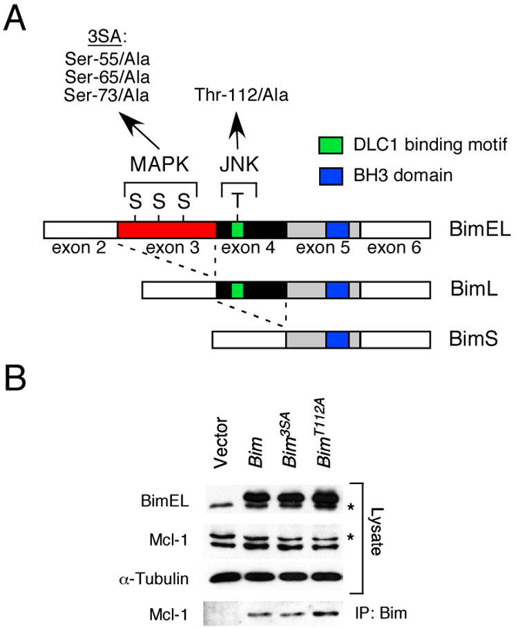
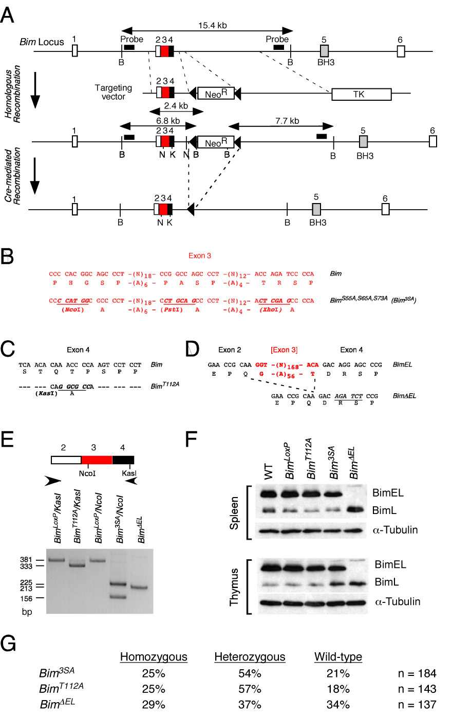
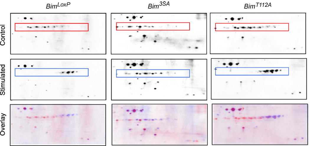
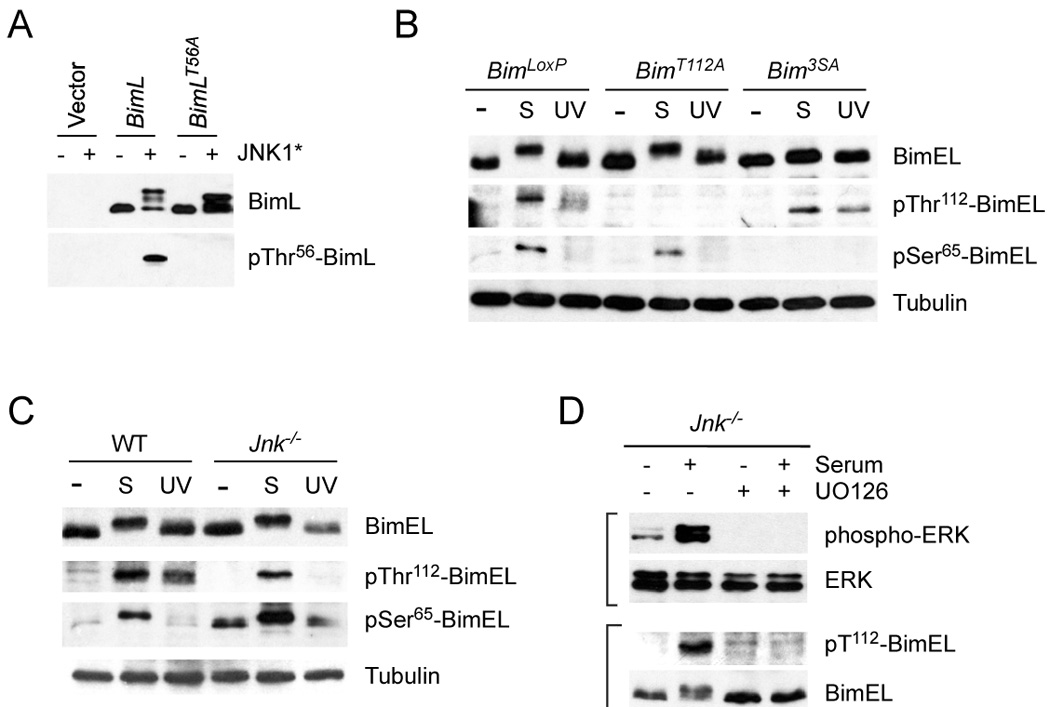
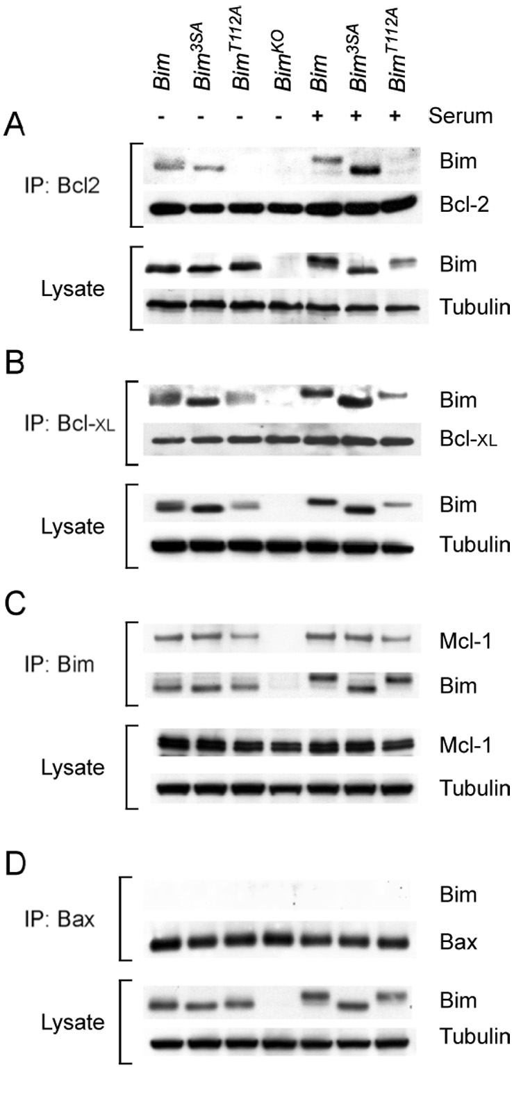
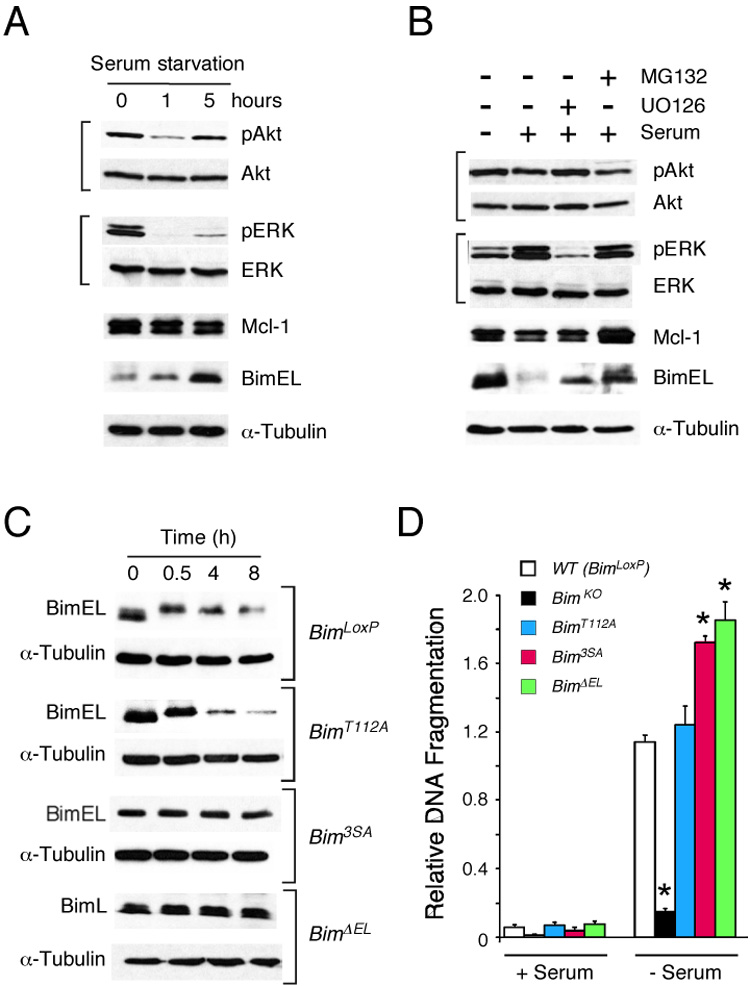
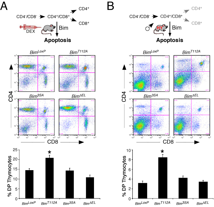
The proapoptotic BH3-only protein Bim is established to be an important mediator of signaling pathways that induce cell death. Multisite phosphorylation of Bim by several members of the MAP kinase group is implicated as a regulatory mechanism that controls the apoptotic activity of Bim. To test the role of Bim phosphorylation in vivo, we constructed mice with a series of mutant alleles that express phosphorylation-defective Bim proteins. We show that mutation of the phosphorylation site Thr-112 causes decreased binding of Bim to the antiapoptotic protein Bcl2 and can increase cell survival. In contrast, mutation of the phosphorylation sites Ser-55, Ser-65, and Ser-73 can cause increased apoptosis because of reduced proteasomal degradation of Bim. Together, these data indicate that phosphorylation can regulate Bim by multiple mechanisms and that the phosphorylation of Bim on different sites can contribute to the sensitivity of cellular apoptotic responses.


Figures

Figure 1
Figure 1. Phosphorylation of Bim isoforms
A) The structure of BimEL, which is encoded by sequences derived from exons 2–6 of the Bim gene, is illustrated schematically. Alternative splicing can delete sequences derived from exon 3 (BimL) or exons 3 & 4 (BimS) and creates two additional Bim isoforms. Three sites of MAPK phosphorylation are encoded by exon 3 (Ser-55, Ser-65, and Ser-73) and one site of JNK phosphorylation is encoded by exon 4 (Thr-112). Mutation of the three sites of Ser phosphorylation (3SA) and the site of Thr phosphorylation (Thr112A) by replacement with Ala residues is indicated. The BH3 domain and the dynein light chain 1 (DLC1) binding motif are illustrated. B) Recombinant BimEL proteins with mutations at the three sites of MAPK phosphorylation (3SA) or the JNK phosphorylation site (Thr-112) were expressed in 293T cells. The endogenous (asterisk) and recombinant BimEL proteins were detected by immunoblot analysis. Endogenous Mcl-1 (asterisk indicates a non-specific band) and α-Tubulin were also examined. Bim/Mcl-1 complexes were examined by immunoprecipitation (IP) of recombinant Bim proteins with an antibody to the Flag epitope-tag and immunoblot analysis with an antibody to Mcl-1.
Figure 2
Figure 2. Construction of mice with phosphorylation-defective Bim
A) Strategy for construction of mice with phosphorylation-defective Bim proteins. The structure of the Bim genomic locus and the targeting vectors that were employed to create germ-line mutations in exons 3 and 4 are illustrated. B–D) The point mutations in exon 3 (B) and exon 4 (C) are illustrated. The strategy for deletion of exon 3 is also illustrated (D). E) PCR analysis of mouse genomic DNA isolated from BimLoxP mice (wild-type Bim) and from mice with homozygous mutations: BimT112A; Bim3SA; and BimΔEL. The presence of the T112A (KasI) and 3SA (NcoI) mutations were detected by digestion with the indicated restriction enzymes. F) Splenocytes and thymocytes were isolated from wild-type C57BL/6J mice and mice with homozygous modifications in the Bim gene: BimLoxP, BimT112A; Bim3SA; and BimΔEL. The expression of Bim and α-Tubulin was examined by immunoblot analysis. G) Mice obtained from heterozygous crosses of Bim mutant mice were genotyped. The number of homozygous, heterozygous, and wild-type mice are presented as the percentage of the total number (n) of mice.
Figure 3
Figure 3. Analysis of Bim phosphorylation in vivo
Homozygous BimLoxP, Bim3SA, and BimT112A primary MEF were serum-starved (Control) or treated with 10% fetal bovine serum (Stimulated) for 30 mins. Extracts were prepared from the cells and examined by 2D gel electrophoresis (isoelectric focusing (pI range 4 – 7) in the horizontal dimension and SDS-PAGE in the vertical dimension) and immunoblot analysis by probing with an antibody to Bim. Proteins corresponding to isoforms of BimEL are indicated with a box. The Control (red) and Stimulated (blue) images were superimposed using Adobe Photoshop CS3 software (Overlay).
Figure 4
Figure 4. Phosphorylation of Bim on Ser65 and Thr112 in vivo
A) Characterization of an antibody to the Bim phosphorylation site Thr112 on BimEL (Thr56 on BimL). 293T cells were transfected with an empty expression vector or a vector that expresses BimL. The expression of BimL and phosphoThr56-BimL was detected by immunoblot analysis of cells extracts. The effect of co-expression of constitutively activated JNK1 (JNK1*) and the replacement of Thr56 with an Ala residue is shown. B) Homozygous BimLoxP, BimT112A, and Bim3SA primary MEF were serum-starved or treated with serum (S) (10% fetal bovine serum; 30 mins) or UV light (60 J/m2; 60 mins). Cell extracts were examined using antibodies to Bim, phospho-Thr112-Bim, phosphoSer65-Bim, and Tubulin. C) Wild-type and Jnk1−/− Jnk2−/− (Jnk−/−) fibroblasts were serum-starved or treated with serum (S) or UV light. Cell extracts were examined using antibodies to Bim, phospho- Thr112-Bim, phosphoSer65-Bim, and Tubulin. The basal phosphorylation of BimEL on Ser65 was slightly increased in JNK-deficient fibroblasts compared with wild-type fibroblasts. D) Jnk1−/− Jnk2−/− (Jnk−/−) fibroblasts were serum-starved or treated with serum and/or the MEK inhibitor U0126 (30 mins). Cell extracts were examined using antibodies to phospho-ERK, ERK, phospho-Thr112-Bim, and Bim.
Figure 5
Figure 5. Interaction of Bim with Bcl2 family proteins
A) Homozygous BimLoxP, BimT112A, and Bim3SA primary MEF were serum-starved or treated with 10% fetal bovine serum (30 mins). The interaction of Bim with Bcl2 was examined by immunoprecipitation (IP) of cell extracts with an antibody to Bcl2 and examination of the amount of co-immunoprecipitated Bim and Bcl2 by immunoblot analysis. The amount of Bim and Tubulin in the cell lysate was probed by immunoblot analysis. B) The interaction of Bim with Bcl- XL was examined by immunoprecipitation of cell extracts with an antibody to Bcl- XL and examination of the amount of co-immunoprecipitated Bcl- XL and Bim by immunoblot analysis. The amount of Bim and Tubulin in the cell lysate was probed by immunoblot analysis. C) The interaction of Bim with Mcl-1 was examined by immunoprecipitation of cell extracts with an antibody to Bim and examination of the amount of co-immunoprecipitated Mcl-1 and Bim by immunoblot analysis. The amount of Mcl-1 and Tubulin in the cell lysate was probed by immunoblot analysis D) The interaction of Bim with Bax was examined by immunoprecipitation of cell extracts with an antibody to Bax and examination of the amount of co-immunoprecipitated Bax and Bim by immunoblot analysis. The amount of Bim and Tubulin in the cell lysate was probed by immunoblot analysis
Figure 6
Figure 6. The BimEL specific domain encoded by exon 2 is required for MAPK-induced Bim degradation
A) Serum starvation induces the expression of BimEL. Wild-type primary MEF were transferred to serum-free medium. Cell extracts were prepared at 0, 1, and 5 h following serum withdrawal. Protein expression was examined by immunoblot analysis. B) Serum addition causes BimEL degradation. Wild-type primary MEF serum-starved (12 h) and then transferred to fresh medium supplemented with 10% FBS. Cell extracts were prepared at 0 and 8 hrs following serum treatment. The effect of pre-incubation (30 mins) with 10 µM U0126 or 20 µM MG132 was examined. Protein expression was examined by immunoblot analysis. C) Primary MEF were prepared from embryos that express wild-type Bim (BimLoxP) and also from homozygous mutants embryos (BimT112A, Bim3SA, and BimΔEL). The cells were transferred to fresh medium supplemented with 10% fetal bovine serum plus 100 µg/ml cycloheximide. Protein expression was examined by immunoblot analysis. D) Primary MEF were incubated in medium supplemented without and with 10% FBS (25 h.). Apoptotic DNA fragmentation was examined by ELISA. The effect of serum withdrawal to cause apoptosis of MEF that express wild-type Bim (BimLoxP) and also from homozygous mutants embryos (BimKO, BimT112A, Bim3SA, and BimΔEL) is presented as mean relative DNA fragmentation ± SD (n = 4). A statistically significant difference (P < 0.05) in DNA fragmentation compared with BimLoxP MEF is indicated with an asterisk.
Figure 7
Figure 7. Effect of phosphorylation-defective Bim on thymocyte apoptosis in vivo
A) Mice (8 – 12 weeks old) were injected with dexamethasone (8.3 µg / g body mass) or solvent (saline). Thymocytes were isolated at 20 h post-injection and the CD4+ and CD8+ sub-populations were examined by flow cytometry. Representative FACS profiles of thymocytes from dexamethasone-treated homozygous BimLoxP, BimT112A, Bim3SA, and BimΔEL mice are illustrated. The data obtained from groups of 8 –12 mice per genotype are presented as the mean ± SD in the lower panel. A statistically significant increase (t-test; P < 0.05) in the survival of double positive (DP) CD4+CD8+ thymocytes from Bim mutant mice compared with mice that express wild-type Bim (BimLoxP) is indicated with an asterisk. B) The thymocytes of male HY-TCR positive mice (5 – 8 weeks old) on genetic backgrounds that are homozygous for the wild-type Bim protein (BimLoxP) or a phosphorylation-defective Bim protein (BimT112A, Bim3SA, and BimDEL) were examined by flow cytometry. Representative FACS profiles of thymocytes are illustrated. The data obtained from groups of 13 –14 mice per genotype are presented as the mean ± SD in the lower panel. A statistically significant increase (P < 0.05) in the survival of double positive (DP) CD4+CD8+ thymocytes from Bim mutant mice compared with mice that express wild-type Bim (BimLoxP) is indicated with an asterisk.

References

    1. Akiyama T, Bouillet P, Miyazaki T, Kadono Y, Chikuda H, Chung UI, Fukuda A, Hikita A, Seto H, Okada T, et al. Regulation of osteoclast apoptosis by ubiquitylation of proapoptotic BH3-only Bcl-2 family member Bim. Embo J. 2003;22:6653–6664. - PMC - PubMed
    1. Becker EB, Howell J, Kodama Y, Barker PA, Bonni A. Characterization of the c-Jun N-terminal kinase-BimEL signaling pathway in neuronal apoptosis. J Neurosci. 2004;24:8762–8770. - PMC - PubMed
    1. Bouillet P, Cory S, Zhang LC, Strasser A, Adams JM. Degenerative disorders caused by Bcl-2 deficiency prevented by loss of its BH3-only antagonist Bim. Dev Cell. 2001a;1:645–653. - PubMed
    1. Bouillet P, Metcalf D, Huang DC, Tarlinton DM, Kay TW, Kontgen F, Adams JM, Strasser A. Proapoptotic Bcl-2 relative Bim required for certain apoptotic responses, leukocyte homeostasis, and to preclude autoimmunity. Science. 1999;286:1735–1738. - PubMed
    1. Bouillet P, Purton JF, Godfrey DI, Zhang LC, Coultas L, Puthalakath H, Pellegrini M, Cory S, Adams JM, Strasser A. BH3-only Bcl-2 family member Bim is required for apoptosis of autoreactive thymocytes. Nature. 2002;415:922–926. - PubMed

Publication types

MeSH terms