. 2008 May 23;30(4):507-18.
doi: 10.1016/j.molcel.2008.03.011.

Decoding of methylated histone H3 tail by the Pygo-BCL9 Wnt signaling complex

Affiliations

Decoding of methylated histone H3 tail by the Pygo-BCL9 Wnt signaling complex

Marc Fiedler et al. Mol Cell. .

Abstract

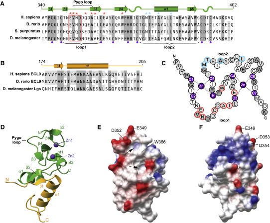
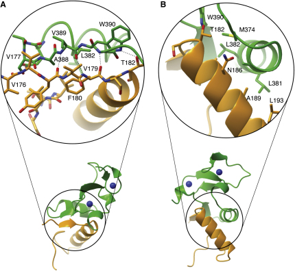
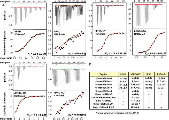
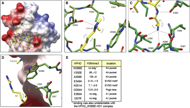
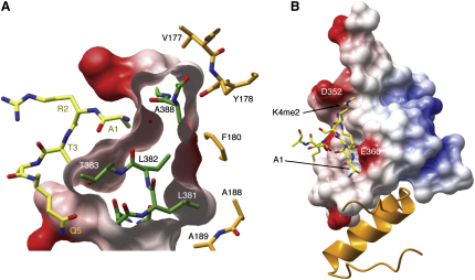
Pygo and BCL9/Legless transduce the Wnt signal by promoting the transcriptional activity of beta-catenin/Armadillo in normal and malignant cells. We show that human and Drosophila Pygo PHD fingers associate with their cognate HD1 domains from BCL9/Legless to bind specifically to the histone H3 tail methylated at lysine 4 (H3K4me). The crystal structures of ternary complexes between PHD, HD1, and two different H3K4me peptides reveal a unique mode of histone tail recognition: efficient histone binding requires HD1 association, and the PHD-HD1 complex binds preferentially to H3K4me2 while displaying insensitivity to methylation of H3R2. Therefore, this is a prime example of histone tail binding by a PHD finger (of Pygo) being modulated by a cofactor (BCL9/Legless). Rescue experiments in Drosophila indicate that Wnt signaling outputs depend on histone decoding. The specificity of this process provided by the Pygo-BCL9/Legless complex suggests that this complex facilitates an early step in the transition from gene silence to Wnt-induced transcription.


Figures

Figure 1
Figure 1
Sequence Alignments and Structure of the Human PHD-HD1 Complex (A and B) Alignments of (A) PHD sequences of hPygo1 (Q9Y3Y4), zebrafish Pygo2 (Q1L8T6), sea urchin Pygo (XP_791313), and Drosophila Pygo (Q9V9W8), and (B) HD1 sequences of hBCL9 (O00512), zebrafish BCL9 (Q67FY0), and Drosophila Lgs (Q961D9). Dark gray, invariant residues; light gray, semiconserved residues. Marked above sequences are secondary structure elements (β sheets, α helices; α turns are marked by S shapes) and residues involved in HD1 (blue) or H3K4me binding (red) as defined by mutational analysis (Figures 3B and 4E). EVND motif is boxed; indicated are also Zn2+-coordinating residues (purple), Pygo loop, and loop1 and loop2 surfaces (brackets). (C) Crossbrace ligation of hPygo1 PHD finger, with Zn-coordinating and mutated residues highlighted (colors as in [A]). (D) Ribbon representation of the hPHD-HD1 complex structure solved at 1.59 Å resolution, with secondary structure elements labeled as in (A) and (B). PHD, green; HD1, orange; Zn2+, purple. (E and F) Molecular surface representations of hPHD-HD1, colored according to electrostatic potential (red, negative charges; blue, positive charges), with some Pygo loop and EVND residues labeled. (E) View similar to (D), showing the PHD loop1 surface, with two conspicuous cavities (arrows) separated by W366; (F) is rotated 180° with respect to (E).
Figure 2
Figure 2
The Pygo-BCL9 Interface Contacts between PHD (green) and HD1 (orange). (A) Parallel β sheets (PHD β5, HD1 β1), (B) α helices. H bonds are indicated by dotted lines; amino acids involved in intermolecular interactions are depicted in cylinder mode.
Figure 3
Figure 3
Binding Affinities between Histone Peptides and PHD or PHD-HD1 (A) ITC profiles for the binding of methylated and unmodified 15-mer histone H3 tail peptides to free hPHD, hPHD-HD1, or dPHD-HD1 complex, as indicated in the panels; data were fitted to a one-site model. Kd values are given in the individual panels (with fitting errors indicated; see the Supplemental Experimental Procedures). (B) Binding constants (Kd values in μM) of free human and Drosophila PHD finger versus PHD-HD1 complex for various histone ligands, as indicated.
Figure 4
Figure 4
Structures of the Ternary Complex, and H3K4me-Binding Cavities (A) Molecular surface representation of hPHD-HD1 binding to H3K4me2 (in yellow cylinder style), with W366 and other critical residues labeled. (B and C) Cylinder representations of (B) semiaromatic K4me2 cavity and (C) A1 cavity, with critical H bonds indicated as dotted lines and hydrophobic contacts as double brackets. (D) Molecular surface representation of PHD, revealing solvent exposure of R2 (regardless of its methylation status). H3K4me, yellow; PHD cavity residues, green. Note that Tern2 has essentially the same structure as Tern1 (shown here; see text). (E) Binding constants of various hPHD point mutants for H3K4me3 15-mer (Kd values in μM; see also Figure 3).
Figure 5
Figure 5
Buttressing of the PHD A1 Cavity by HD1 (A) Molecular surface representation of PHD (green) with electrostatic potential, facing A1 cavity (left, yellow) and HD1 (right, orange). (B) Buttressing of A1 cavity of PHD (molecular surface representation with electrostatic potential) by HD1 (ribbon representation). D352 lip residue of the K4me2 cavity and E360 residue critical for A1 anchoring are indicated.
Figure 6
Figure 6
Rescue Assays in Drosophila, Revealing Functional Relevance of K4me Cavity (A–C) Wing discs of third instar larvae bearing pygoS28 mutant clones (marked by absence of GFP), with or without overexpressed HA-Pygo, double stained with (A) DAPI (to reveal the nuclei) or (B and C) anti-HA antibody, and (A–C) anti-Senseless antibodies. (D–F) Anterior wing margin of adult flies bearing pygoS28 mutant clones, with or without overexpressed HA-Pygo. (A and D) controls, (B and E) WT HA-Pygo, (C and F) HA-Pygo-V757E.

References

    1. Adachi S., Jigami T., Yasui T., Nakano T., Ohwada S., Omori Y., Sugano S., Ohkawara B., Shibuya H., Nakamura T., Akiyama T. Role of a BCL9-related β-catenin-binding protein, B9L, in tumorigenesis induced by aberrant activation of Wnt signaling. Cancer Res. 2004;64:8496–8501. - PubMed
    1. Arce L., Yokoyama N.N., Waterman M.L. Diversity of LEF/TCF action in development and disease. Oncogene. 2006;25:7492–7504. - PubMed
    1. Belenkaya T.Y., Han C., Standley H.J., Lin X., Houston D.W., Heasman J., Lin X. pygopus encodes a nuclear protein essential for wingless/Wnt signaling. Development. 2002;129:4089–4101. - PubMed
    1. Bienz M. The PHD finger, a nuclear protein-interaction domain. Trends Biochem. Sci. 2006;31:35–40. - PubMed
    1. Bienz M., Clevers H. Linking colorectal cancer to Wnt signaling. Cell. 2000;103:311–320. - PubMed

Publication types

MeSH terms