Review
. 2008 Jun;34(6):645-51.
doi: 10.1016/j.joen.2008.03.001.

The hidden treasure in apical papilla: the potential role in pulp/dentin regeneration and bioroot engineering

Affiliations
Review

The hidden treasure in apical papilla: the potential role in pulp/dentin regeneration and bioroot engineering

George T-J Huang et al. J Endod. 2008 Jun.

Abstract

Some clinical case reports have shown that immature permanent teeth with periradicular periodontitis or abscess can undergo apexogenesis after conservative endodontic treatment. A call for a paradigm shift and new protocol for the clinical management of these cases has been brought to attention. Concomitantly, a new population of mesenchymal stem cells residing in the apical papilla of permanent immature teeth recently has been discovered and was termed stem cells from the apical papilla (SCAP). These stem cells appear to be the source of odontoblasts that are responsible for the formation of root dentin. Conservation of these stem cells when treating immature teeth may allow continuous formation of the root to completion. This article reviews current findings on the isolation and characterization of these stem cells. The potential role of these stem cells in the following respects will be discussed: (1) their contribution in continued root maturation in endodontically treated immature teeth with periradicular periodontitis or abscess and (2) their potential utilization for pulp/dentin regeneration and bioroot engineering.


Figures

Figure 1
Figure 1
Apical papilla. (A) An extracted human third molar depicting three immature roots with two pieces of apical papilla being removed from their apices (arrow heads) and one piece of apical papilla being peeled away from the root end but not completely detached (arrow). (B) A developing root tip with attached apical papilla was cultured in vitro for 3 days before being processed for hematoxylin and eosin (H&E) staining. Odontoblasts (black arrows), apical cell-rich zone (open arrowheads), and apical papilla tissue are indicated. (C) Magnified view of the area indicated by the yellow rectangle.
Figure 2
Figure 2
The potential role of apical papilla in root maturation in human and minpig. (A) Human incisor crown fracture (arrows) occurred before the completion of root development. (B) Immediate root canal treatment was performed on the fractured tooth. Pulp tissue was completely removed and root canal was sealed with filling material. (C) Three months after endodontic treatment, the root tip continued to develop. (D) Seven months after treatment, a significant amount of root tip was formed. (E) When distal buccal root apical papilla of the lower first molar was surgically removed from a 9-month-old minipig, the distal buccal root stopped developing at the 3-month follow-up (black arrows), but other roots show a normal growth and development (red arrows).
Figure 3
Figure 3
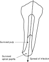
The hypothetical pathway of infection of immature permanent teeth. The infection may pass through the survived pulp and apical papilla reaching the periradicular tissues and causing extensive bone resorption.
Figure 4
Figure 4
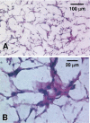
DPSCs grown in PLG in vitro. DPSCs were seeded onto PLG scaffolds cylindrical shape, 5 (diameter) × 2 (height) mm with pore diameters between 250 and 425 μm kindly provided by Dr L.D. Shea (Northwestern University, Evanston, IL) and cultured in vitro for 7 weeks. Cells attached onto the inner surfaces of the scaffold (H&E). The scaffolds without cells (control, not shown) were also processed for H&E, and no visible staining was observed (original magnification: A, ×100; B, ×400).
Figure 5
Figure 5
A schematic illustration of proposed stepwise insertion of engineered pulp tissue in the clinical setting.

References

    1. Iwaya SI, Ikawa M, Kubota M. Revascularization of an immature permanent tooth with apical periodontitis and sinus tract. Dental Traumatol. 2001;17:185–7. - PubMed
    1. Banchs F, Trope M. Revascularization of immature permanent teeth with apical periodontitis: new treatment protocol? J Endod. 2004;30:196–200. - PubMed
    1. Chueh L-H, Huang GTJ. Immature teeth with periradicular periodontitis or abscess undergoing apexogenesis: A paradigm shift. J Endod. 2006;32:1205–13. - PubMed
    1. Sonoyama W, Liu Y, Fang D, et al. Mesenchymal stem cell-mediated functional tooth regeneration in Swine. PLoS ONE. 2006;1:e79. - PMC - PubMed
    1. Sonoyama W, Liu Y, Yamaza T, et al. Characterization of the apical papilla and its residing stem cells from human immature permanent teeth: a pilot study. J Endod. 2008;34:166–71. - PMC - PubMed

Publication types

MeSH terms