Review
. 2008 Jul;32(5):1024-43.
doi: 10.1016/j.neubiorev.2008.04.002. Epub 2008 Apr 18.

The taste of sugars

Affiliations
Review

The taste of sugars

Stuart A McCaughey. Neurosci Biobehav Rev. 2008 Jul.

Abstract

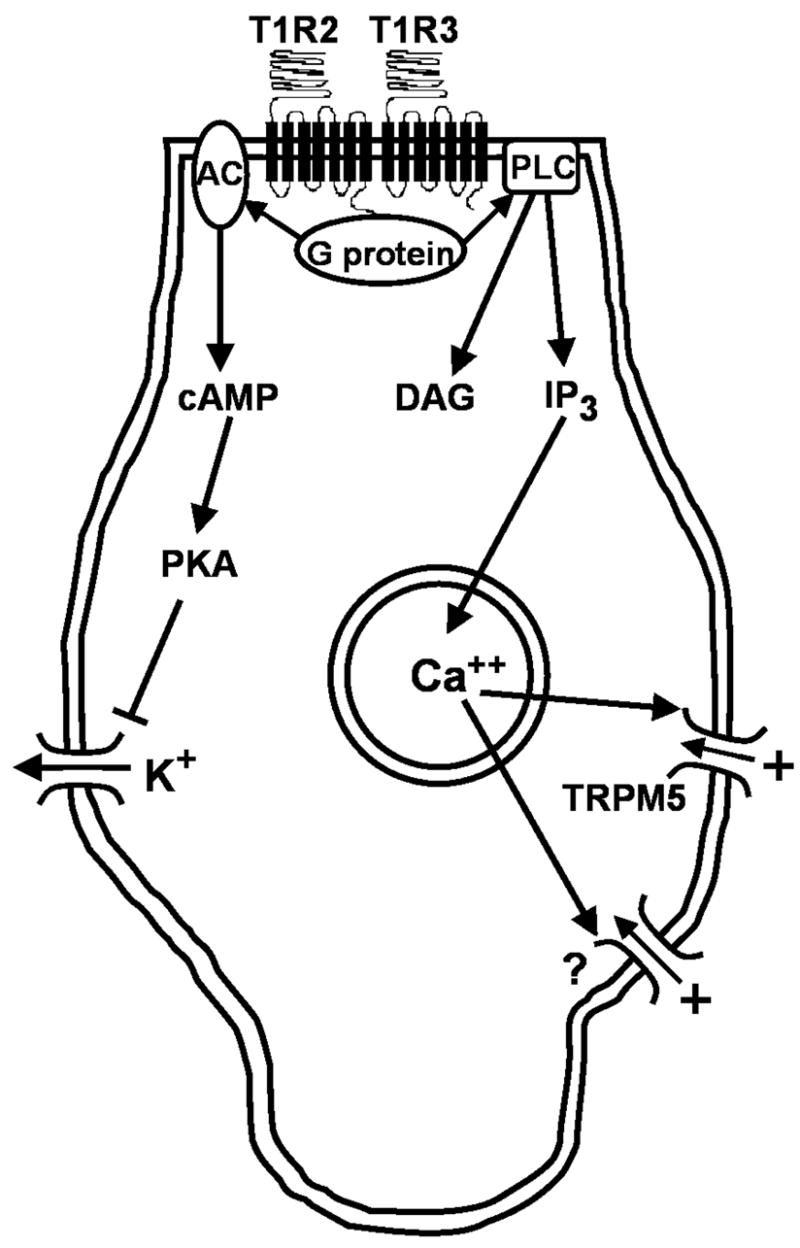
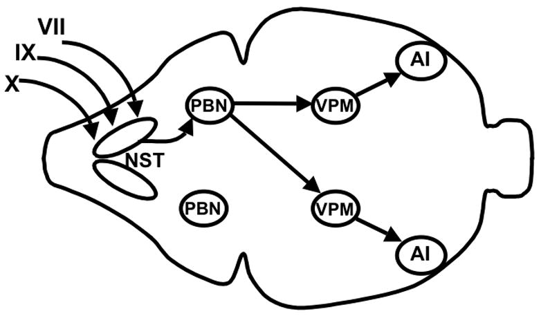
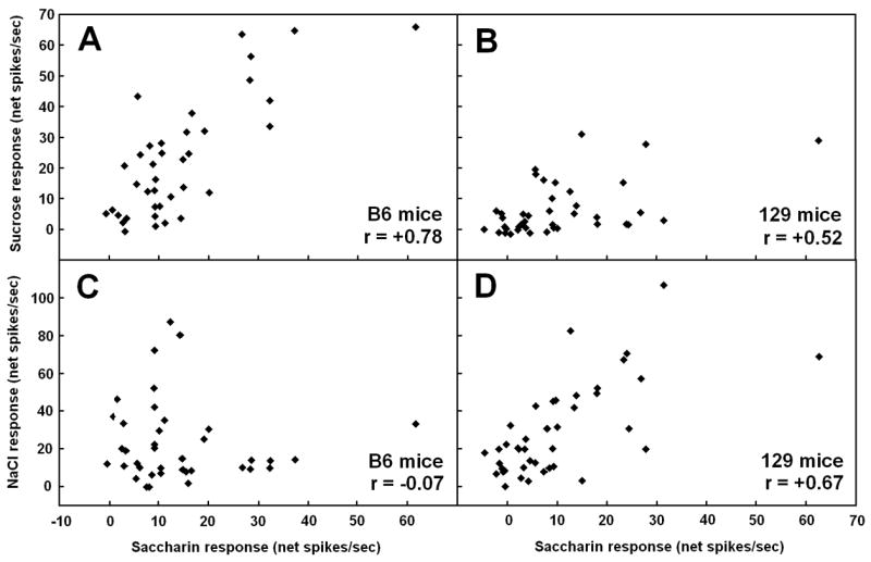
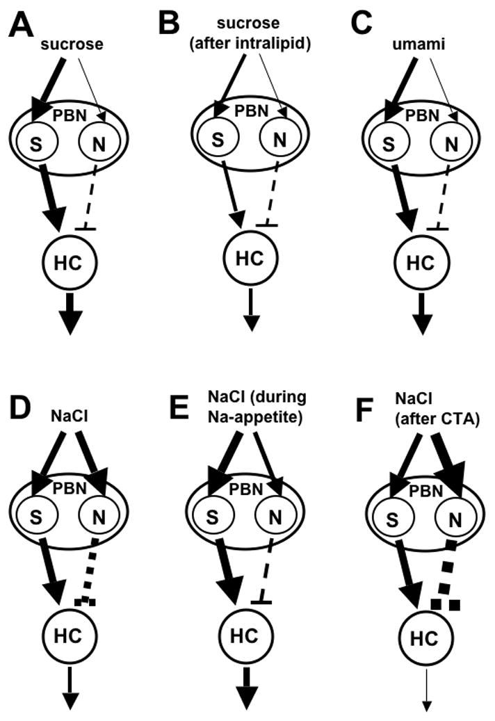
Sugars evoke a distinctive perceptual quality ("sweetness" in humans) and are generally highly preferred. The neural basis for these phenomena is reviewed for rodents, in which detailed electrophysiological measurements have been made. A receptor has been identified that binds sweeteners and activates G-protein-mediated signaling in taste receptor cells, which leads to changes in neural firing rates in the brain, where perceptions of taste quality, intensity, and palatability are generated. Most cells in gustatory nuclei are broadly tuned, so quality perception presumably arises from patterns of activity across neural populations. However, some manipulations affect only the most sugar-oriented cells, making it useful to consider them as a distinct neural subtype. Quality perception may also arise partly due to temporal patterns of activity to sugars, especially within sugar-oriented cells that give large but delayed responses. Non-specific gustatory neurons that are excited by both sugars and unpalatable stimuli project to ventral forebrain areas, where neural responses provide a closer match with behavioral preferences. This transition likely involves opposing excitatory and inhibitory influences by different subgroups of gustatory cells. Sweeteners are generally preferred over water, but the strength of this preference can vary across time or between individuals, and higher preferences for sugars are often associated with larger taste-evoked responses.


Figures

Figure 1
Figure 1
Intracellular cascades that follow binding of sugars to a putative taste receptor and that result in depolarization of a taste receptor cell. Sugars are thought to bind to a dimer of the proteins T1R2 and T1R3. T1R2 then activates a complex of G proteins which contains alpha, beta, and gamma subunits, including α-gustducin in some cases. One transduction pathway involves G-protein-mediated activation of adenylate cyclase (AC), which then increases production of cyclic AMP (cAMP), which causes inhibition of basolateral K+ channels through PKA. Depolarization of the cell results from decreased K+ efflux. An alternate pathway may also be involved, though evidence is stronger for artificial sweeteners than for sugars. This pathway involves activation of phospholipase C B2 (PLC) by the T1R2-coupled G-protein. PLC then causes inositol triphosphate (IP3) to release Ca++ from intracellular stores. Calcium opens non-selective TRPM5 channels, which allow cations to enter the cell, causing depolarization. There is also evidence for an unidentified cation channel (?) that is activated by Ca++ and can induce depolarization of the cell.
Figure 2
Figure 2
Schematic overhead view of the main central gustatory pathways in the rat. Projections originating from only one side of the oral cavity are shown for the sake of simplicity. Branches of the facial (VII), glossopharyngeal (IX), and vagus (X) nerves innervate taste buds in the tongue and other parts of the oral cavity. These peripheral taste-responsive fibers synapse in the nucleus of the solitary tract (NST) in the caudal hindbrain. Projections are then sent rostrally to the ipsilateral parabrachial nucleus (PBN), which projects bilaterally to the ventral posterior medial subnucleus of the thalamus (VPM). The VPM then projects ipsilaterally to the cortical gustatory area in agranular insular cortex (AI).
Figure 3
Figure 3
Comparison of gustatory stimuli based on activity in the nucleus of the solitary tract of C57BL/6ByJ mice. Across-neuron patterns of activity evoked by stimuli were correlated with each other, and multidimensional scaling was used to generate a two-dimensional space in which stimuli with similar patterns were located close to each other. The sugars sucrose and maltose are grouped with other compounds that are thought to taste sweet to mice and in a distinct location from compounds thought to taste salty, sour, or bitter. ACE, 20 mM acesulfame-K; CA, 100 mM CaCl2; CI, 10 mM citric acid; GLY, 100 mM glycine; HCL, 10 mM HCl; IMP, 10 mM disodium inosine 5′ monophosphate; MAL, 500 mM maltose; NA, 100 mM NaCl; NH, 100 mM NH4Cl; PHE, 100 mM D-phenylalanine; PRO, 100 mM L-proline; Q, 20 mM quinine HCl; SAC, 10 mM NaSaccharin; SC, 1 mM SC-45647; SUC, 500 mM sucrose. Reprinted from McCaughey, 2007 with permission.
Figure 4
Figure 4
Sweeteners evoke independent phasic and tonic responses in the nucleus of the solitary tract (NST) of C57BL/6ByJ (B6) mice. A–C. Post-stimulus time histograms showing mean net responses to three sweeteners in different subgroups of NST cells during the first 2.5 sec after stimulus onset in 100-ms bins. Sucrose at 500 mM (A), 500 mM maltose (B), and the artificial sweetener SC-45647 at 1 mM (C) evoked small responses with a rapid onset in NST cells that had NaCl- or HCl-oriented profiles based on evoked responding across a 5-sec evoked period (n = 17; dashed lines). In contrast, these compounds evoked responses that were larger, but more delayed, in cells with sucrose-oriented response profiles (n = 21; solid lines). D–E. Mean net responses to 500 mM sucrose in the NST of B6 mice during phasic (D) and tonic (E) response periods. Responses that deviated significantly from baseline firing are indicated by solid bars, whereas ones that did not are indicated by open bars. Data are shown for 38 individual neurons, placed in descending order of their phasic response to sucrose for both graphs. The phasic period was defined as the first 600 ms after stimulus onset, and the tonic period lasted from 600–5000 ms after onset. Both periods allowed adequate time for cells to show a significant change in firing rate compared to baseline. However, the Pearson product moment correlation between cells’ phasic and tonic sucrose responses was −0.06, indicating that the two periods are independent. This figure is previously unpublished, but data are taken from the experiment for McCaughey, 2007.
Figure 5
Figure 5
Scatterplots showing the relationship between responses evoked by 10 mM NaSaccharin and those evoked by 500 mM sucrose (A,B) or 100 mM NaCl (C,D) in cells from the nucleus of the solitary tract of C57BL/6ByJ (B6; A,C) or 129P3/J (129; B,D) mice. The Pearson product moment correlation is shown in the bottom right of each panel. Responses to saccharin were more highly correlated with responses to sucrose in B6 mice than in 129 mice, whereas saccharin responses were more highly correlated with NaCl responses in 129 animals than in the B6 strain. These results suggests that saccharin tastes more sweet and less salty to B6 mice than to 129 mice, given that the correlation between across-neuron profiles provides a good match with similarity of perceived taste quality. Such a perceptual difference between strains likely contributes to the higher preferences for saccharin in B6 mice compared to 129 mice. This figure is previously unpublished, but data are taken from the experiment for McCaughey, 2007.
Figure 6
Figure 6
Model to explain how the firing of broadly-tuned gustatory cells could result in post-synaptic responses that closely track the palatability of a taste stimulus. The gustatory signal is sent to a sucrose-oriented cell in the PBN (S), which then excites a hypothetical hedonically-oriented cell in the ventral forebrain (HC), whose responses are related more closely to stimulus palatability than taste quality. Taste stimuli can also drive firing in a NaCl-oriented PBN cell (N), which inhibits the HC. Line thicknesses are proportional to evoked firing rates, and neural excitation is indicated by arrows and inhibition by dashed lines. A) Oral sucrose causes robust responses in PBN S-cells, but weak responses in N-cells, resulting in a large response by the HC. B) Infusion of lipids is known to reduce sucrose intake, as well as the size of sucrose responses in S-cells in the PBN, possibly due to smaller input from the NST, though interactions between taste and visceral inputs in the PBN are another potential source of the effect. PBN N-cell responses are unaffected by lipid infusion. The net result in the HC would be smaller excitation by sucrose. C) Umami stimuli, such as a mixture of MSG and GMP, are known to evoke larger responses in S- than N-cells in the NST and PBN. The summation of PBN outputs would cause excitation of the HC by umami compounds, which tend to be highly preferred. D) PBN S-cells respond not only to sucrose, but also are strongly excited by moderately preferred or avoided concentrations of NaCl. However, NaCl also effectively drives firing of N-cells in the PBN, with the net result that the HC would not give large response to NaCl under normal conditions. E) When rats are sodium-deprived, or when an appetite for NaCl is created by other means, there is a reduction in the size of the response evoked by N-cells in the NST, which provide input to N-cells in the PBN. There is also a report of elevated S-cell responses to NaCl in the NST of sodium-deprived rats. Thus, sodium appetite would be accompanied by robust HC responses when NaCl is tasted. F) Creation of a conditioned taste aversion to NaCl causes a decrease in NaCl intake and palatability in rats. There is also an increase in the taste-evoked response to NaCl in the PBN, which is limited to amiloride-sensitive cells that are narrowly tuned to NaCl. Stronger excitatory input to these PBN N-cells is depicted as a possible cause of this elevated responding to NaCl, though this has not been tested directly.

References

    1. Accolla R, Bathellier B, Petersen CC, Carleton A. Differential spatial representation of taste modalities in the rat gustatory cortex. J Neurosci. 2007;27(6):1396–404. - PMC - PubMed
    1. Ackerman SH, Albert M, Shindledecker RD, Gayle C, Smith GP. Intake of different concentrations of sucrose and corn oil in preweanling rats. Am J Physiol. 1992;262:R624–7. - PubMed
    1. Adler E, Hoon MA, Mueller KL, Chandrashekar J, Ryba NJ, Zuker CS. A novel family of mammalian taste receptors. Cell. 2000;100(6):693–702. - PubMed
    1. Agmo A, Marroquin E. Role of gustatory and postingestive actions of sweeteners in the generation of positive affect as evaluated by place preference conditioning. Appetite. 1997;29(3):269–89. - PubMed
    1. Aleksanyan ZA, Buresova O, Bures J. Modification of unit responses to gustatory stimuli by conditioned taste aversion in rats. Physiol Behav. 1976;17(2):173–9. - PubMed

Publication types