. 2008 Apr-May;78(4-5):293-304.
doi: 10.1016/j.plefa.2008.04.001. Epub 2008 May 21.

The aging human orbitofrontal cortex: decreasing polyunsaturated fatty acid composition and associated increases in lipogenic gene expression and stearoyl-CoA desaturase activity

Affiliations

The aging human orbitofrontal cortex: decreasing polyunsaturated fatty acid composition and associated increases in lipogenic gene expression and stearoyl-CoA desaturase activity

Robert K McNamara et al. Prostaglandins Leukot Essent Fatty Acids. 2008 Apr-May.

Abstract

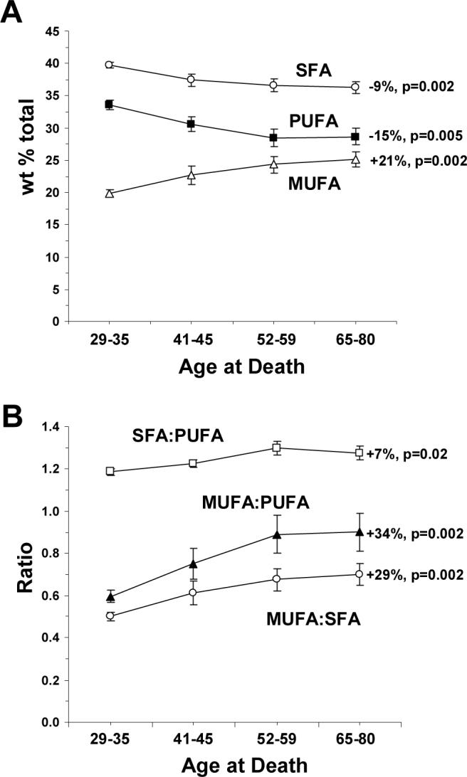
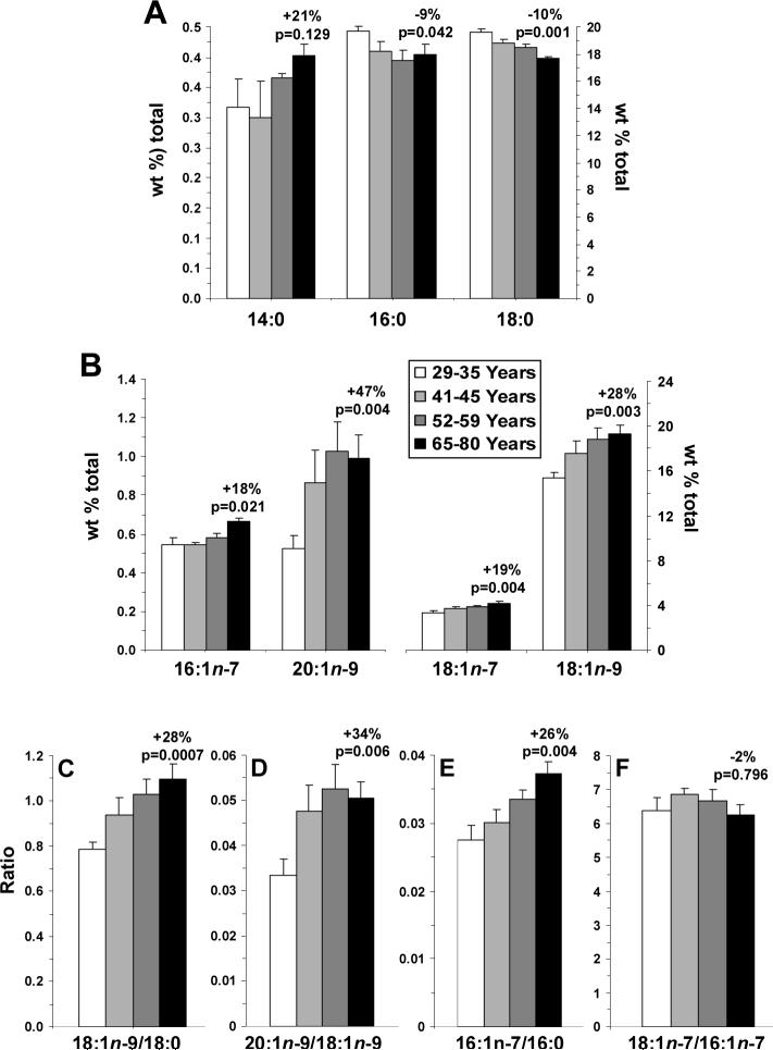
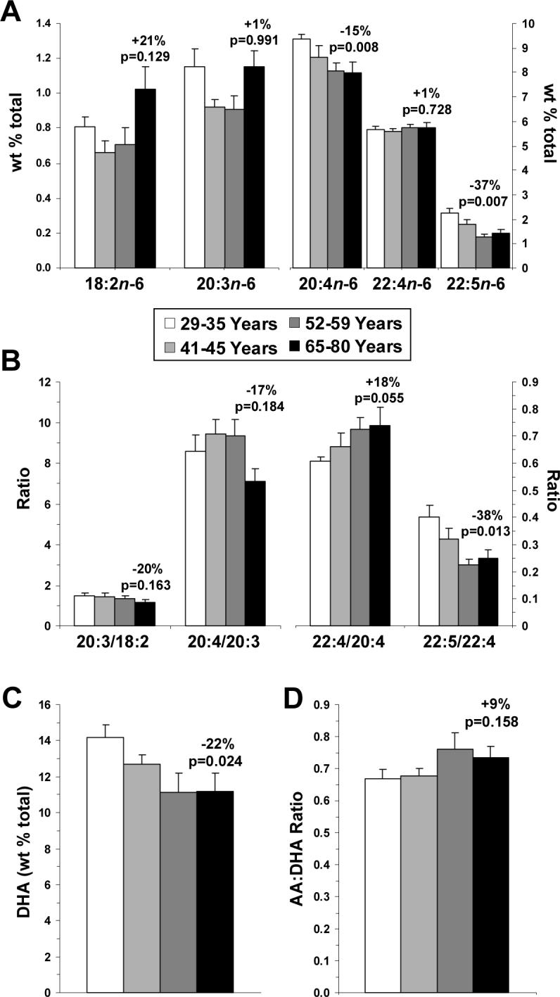
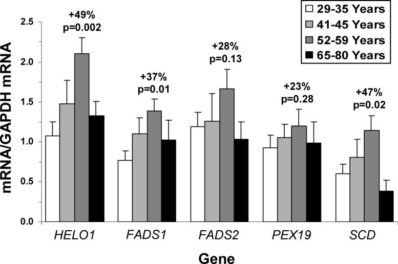
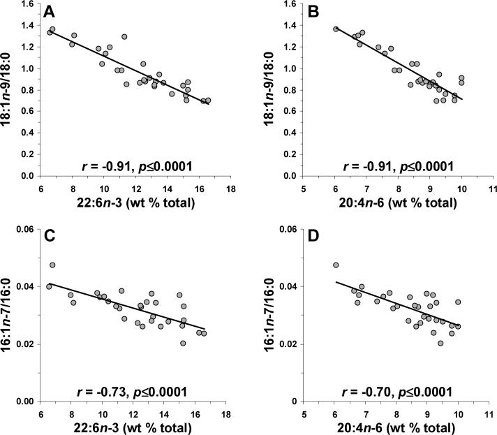
Orbitofrontal cortex (OFC, Brodmann area 10) gray matter volume reductions and selective reductions in docosahexaenoic acid (DHA, 22:6n-3) are observed in adult patients with major depressive disorder (MDD). OFC gray matter volume also decreases with advancing age in healthy subjects. To examine if OFC gray matter DHA composition decreases during normal aging, we determined age-related changes in OFC gray matter fatty acid composition by gas chromatography in subjects aged 29-80 years (n=30). We additionally determined elongase (HELO1), delta-5 desaturase (FASD1), delta-6 desaturase (FASD2), peroxisomal (PEX19), and stearoyl-CoA desaturase (SCD) mRNA expression in the same tissues. Increasing age was associated with a progressive decline in polyunsaturated fatty acid (PUFA) composition, including DHA and arachidonic acid (AA, 20:4n-6), and transient, apparently compensatory, elevations in elongase and desaturase gene expression. The age-related reduction in PUFA composition was inversely correlated with SCD expression and activity resulting in elevations in monounsaturated fatty acid composition. These dynamic age-related changes in OFC gray matter fatty acid composition and biosynthetic gene expression may contribute to the progressive decline in OFC gray matter volume found with advancing age. The implications of age-related reductions in OFC PUFA composition for affective dysregulation in the elderly are discussed.


Figures

Figure 1
Figure 1
Age-related changes in fatty acid class composition (wt % total) in the four age subgroups (29−35 yrs, 41−45 yrs, 52−59 yrs, and 65−80 yrs): (A) Age-related changes in saturated fatty acid (SFA) composition (∑14:0, 16:0, 18:0), monounsaturated fatty acid (MUFA) composition (∑16:1n-7, 18:1n-7, 18:1n-9, 20:1n-9), and polyunsaturated fatty acid (PUFA) composition (∑18:2n-6, 20:3n-6, 20:4n-6, 22:4n-6, 22:5n-6, 22:6n-3). (B) Age-related changes in the ∑MUFA:∑SFA ratio, the ∑SFA:∑PUFA ratio, and the ∑MUFA:∑PUFA ratio in the four age subgroups. Data are expressed as mean ± S.E.M. Percent change in the oldest versus the youngest subgroup and associated p-values (unpaired t-test, two-tailed) are presented.
Figure 2
Figure 2
Age-related changes in individual SFA and MUFA compositions (wt % total) in the four age subgroups (29−35 yrs, 41−45 yrs, 52−59 yrs, and 65−80 yrs): (A) myristic acid (14:0), heptadecanoic acid (17:0), palmitic acid (16:0) and stearic acid (18:0) compositions, (B) palmitioleic acid (16:1n-7), eicosenoic acid (20:1n-9), cis-vaccenic acid (18:1n-7), and oleic acid (18:1n-9) compositions, (C) the 18:1n-9/18:0 ratio, (D) the 20:1n-9/18:1n-9 ratio, (E) the 16:1n-7/16:0 ratio, and (F) the 18:1n-7/16:1n-7 ratio. Data are expressed as mean ± S.E.M. Percent change in the oldest versus the youngest subgroup and associated p-values (unpaired t-test, two-tailed) are presented.
Figure 3
Figure 3
Age-related changes in individual PUFA compositions (wt % total) in the four age subgroups (29−35 yrs, 41−45 yrs, 52−59 yrs, and 65−80 yrs): (A) linoleic acid (18:2n-6), homo-γ1 linoleic acid (20:3n-6), arachidonic acid (AA, 20:4n-6), adrenic acid (22:4n-6), and docosapentaenoic acid (22:5n-6) compositions, (B) 20:3/18:2, 20:4/20:3, 22:4/20:4, and 22:5/22:4 ratios, (C) docosahexaenoic acid (DHA, 22:6n-3) composition, and (D) the AA:DHA ratio. Data are expressed as mean ± S.E.M. Percent change in the oldest versus the youngest subgroup and associated p-values (unpaired t-test, two-tailed) are presented.
Figure 4
Figure 4
Age-related changes in lipogenic mRNA expression in the OFC gray matter of the four age subgroups (29−35 yrs, 41−45 yrs, 52−59 yrs, and 65−80 yrs): HELO1 (elongase), FASD1 (delta-5 desaturase), FASD2 (delta-6 desaturase), PEX19 (peroxisome), and SCD (delta-9 desaturase) mRNA expression. Data are expressed as mean mRNA/GAPDH mRNA ± S.E.M. Percent change from the 52−59 yrs subgroup and associated p-values (unpaired t-test, two-tailed) are presented.
Figure 5
Figure 5
Linear regression analyses of the 18:1n-9:18:0 ratio and DHA (22:6n-3) (A) and AA (20:4n-6) (B) compositions, and the 16:1n-7:16:0 ratio and DHA (C) and AA (D) compositions. Note that both 18:1n-9/18:0 and 16:1n-7/16:0 ratios, indices of stearoyl-CoA desaturase activity, are inversely correlated with both DHA and AA composition. Pearson correlation coefficients and associated p-values (two-tailed) are presented.

References

    1. Kringelbach ML, Rolls ET. The functional neuroanatomy of the human orbitofrontal cortex: evidence from neuroimaging and neuropsychology. Prog Neurobiol. 2004;72:341–372. - PubMed
    1. Raz N, Gunning FM, Head D, Dupuis JH, McQuain J, Briggs SD, Loken WJ, Thornton AE, Acker JD. Selective aging of the human cerebral cortex observed in vivo: differential vulnerability of the prefrontal gray matter. Cereb Cortex. 1997;7:268–282. - PubMed
    1. Raz N, Gunning-Dixon F, Head D, Rodrigue KM, Williamson A, Acker JD. Aging, sexual dimorphism, and hemispheric asymmetry of the cerebral cortex: replicability of regional differences in volume. Neurobiol Aging. 2004;25:377–396. - PubMed
    1. Resnick SM, Pham DL, Kraut MA, Zonderman AB, Davatzikos C. Longitudinal magnetic resonance imaging studies of older adults: a shrinking brain. J Neurosci. 2003;23:3295–3301. - PMC - PubMed
    1. Tisserand DJ, van Boxtel MP, Pruessner JC, Hofman P, Evans AC, Jolles J. A voxel-based morphometric study to determine individual differences in gray matter density associated with age and cognitive change over time. Cereb Cortex. 2004;14:966–973. - PubMed

Publication types