. 2008 Jul 18;283(29):20060-8.
doi: 10.1074/jbc.M802940200. Epub 2008 May 22.

Histone Arg modifications and p53 regulate the expression of OKL38, a mediator of apoptosis

Affiliations

Histone Arg modifications and p53 regulate the expression of OKL38, a mediator of apoptosis

Hongjie Yao et al. J Biol Chem. .

Abstract

Protein Arg methyltransferases function as coactivators of the tumor suppressor p53 to regulate gene expression. Peptidylarginine deiminase 4 (PAD4/PADI4) counteracts the functions of protein Arg methyltransferases in gene regulation by deimination and demethylimination. Here we show that the expression of a tumor suppressor gene, OKL38, is activated by the inhibition of PAD4 or the activation of p53 following DNA damage. Chromatin immunoprecipitation assays showed a dynamic change of p53 and PAD4 occupancy and histone Arg modifications at the OKL38 promoter during DNA damage, suggesting a direct role of PAD4 and p53 in the expression of OKL38. Furthermore, we found that OKL38 induces apoptosis through localization to mitochondria and induction of cytochrome c release. Together, our studies identify OKL38 as a novel p53 target gene that is regulated by PAD4 and plays a role in apoptosis.


Figures

FIGURE 1.
FIGURE 1.
Identification of OKL38 as a gene regulated by PAD4. A and B, effects of Cl-amidine on the expression of OKL38 in MCF-7 cells (A) or U2OS cells (B) were analyzed by qRT-PCR at 24 h after Cl-amidine treatment. The amounts of OKL38 mRNA were normalized with GAPDH. The amount of OKL38 mRNA without Cl-amidine treatment was normalized as 1-fold. Averages and standard deviations are shown (n = 3). C, OKL38 expression was activated by Cl-amidine in a dosage-dependent manner in MCF-7 cells (n = 3). D, amounts of OKL38 protein were analyzed by Western blot before and after 200μm Cl-amidine treatment in MCF-7 or U2OS cells. E, cells were treated by the PAD4 siRNAs or a control green fluorescent protein siRNA (GFP-siRNA) for 3 days. The amounts of PAD4 were analyzed by Western blot. Note the doublet bands of PAD4. F, effects of PAD4 depletion on the expression of OKL38 in U2OS cells were analyzed by qRT-PCR. G, PAD4 association, H3R17 methylation, and H3 citrullination levels at the OKL38 promoter were examined by ChIP in U2OS cells before and after Cl-amidine treatment. Averages of ChIP signals and standard deviations are shown (n = 3). Representative PCR results of the ChIP DNA samples are shown in supplemental Fig. S2. The GAPDH promoter was analyzed as a control.
FIGURE 2.
FIGURE 2.
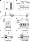
Expression of OKL38 is regulated by p53. A, change of OKL38 gene expression was analyzed by qRT-PCR in the p53–/– H1299 cells before and after the expression of p53. OKL38 expression was normalized to GAPDH (n = 3). B, changes in the OKL38, p21, and GAPDH mRNA levels were monitored by RT-PCR in Tet-On Saos-2 cell lines at 12 h after the addition of doxycycline to induce the expression of wild type p53 or the inactive p53R175H mutant. C, schematic drawing of the promoter region of the OKL38 gene. Exons I and II, intron I, two putative p53-binding sites predicted by the PROMO program were denoted. D, p53 bound the positive control BB9 oligo and the Oligo-1 of the OKL38 promoter, but not Oligo-2, in EMSA. The p53 antibody (PAB421) was used to activate the binding of p53 to its cognate sites in EMSAs. E, competition of p53 binding to the radioactive Oligo-1 or BB9 by extra amount of unlabeled BB9, Oligo-1, and Oligo-2. F, ChIP assays of the binding of p53 to the OKL38 promoter in the p53–/– H1299 cells without or with the ectopic expression of FLAG-p53. G, as a control, p53 was not associated with the GAPDH promoter without or with the expression of FLAG-p53.
FIGURE 3.
FIGURE 3.
OKL38 is inducible by DNA damage and its promoter is regulated by dynamic p53 and PAD4 binding and histone Arg modifications. A and B, changes of OKL38 expression detected by qRT-PCR after doxorubicin treatment in MCF-7 cells (A) or U2OS cells (B). The expression of OKL38 was normalized with that of GAPDH. OKL38 expression at 0 h was normalized as 1. Averages and standard deviations are shown (n = 3). C, levels of the OKL38 expression were examined by qRT-PCR in the p53+/+ and p53–/– HCT116 cells before and after doxorubicin treatment. The expression of OKL38 in the untreated p53+/+ HCT116 cells was normalized as 1. Averages and standard deviations are shown (n = 3). D, dynamic changes of p53, PAD4, histone H3R17 methylation, and H3 citrullination on the OKL38 promoter were analyzed by ChIP analyses in MCF-7 cells. Representative PCR results of the ChIP DNA samples are shown in supplemental Fig. S4, D and E. The amounts of ChIP signals were quantified using the NIH image J program, and signals at 0 h were normalized as 1. Averages and standard deviations are shown (n = 3). The GAPDH promoter was analyzed as a control.
FIGURE 4.
FIGURE 4.
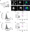
OKL38 overexpression induces apoptosis in U2OS cells. A, FLAG-OKL38 expression was detected at 24 h after transient transfection. Actin was blotted to show equal protein loading. B, expression of OKL38 led to the condensation of the nuclear DNA compared with surrounding cells without FLAG-OKL38 expression. C, induction of the apoptotic body-like structure formation after the overexpression of FLAG-OKL38. White circle outlines a singular nucleus. D and E, flow cytometry analyses of cells transfected with the pIRES vector and stained with annexin V (D) or both annexin V and propidium iodide (PI)(E). F and G, flow cytometry analyses of cells transfected with the FLAG-OKL38/pIRES plasmid and stained with annexin V (F) or both annexin V and propidium iodide (G).
FIGURE 5.
FIGURE 5.
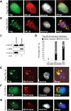
Subcellular localization of OKL38 to nucleus and mitochondria in U2OS cells. A, nuclear localization of FLAG-OKL38 staining was detected in U2OS cells at 24 h after transfection. B, spotted cytoplasmic FLAG-OKL38 staining was also detected in U2OS cells at 24 h after transfection. C, proteins from U2OS cells expressing FLAG-OKL38 were separated into nuclear and cytoplasmic fractions. The presence of FLAG-OKL38, NPM, and tubulin in the nuclear or cytosolic fractions was analyzed by Western blot. D, percentages of cells with mainly cytosolic OKL38 (dark bars) or nuclear OKL38 (gray bars) were analyzed under the microscope. Over 200 U2OS cells expressing FLAG-OKL38 plasmid were counted at each time points (n = 3, standard deviations were shown). E, double staining of U2OS cells with the FLAG antibody and a mitochondria dye, MitoTracker, showed the localization of FLAG-OKL38 in mitochondria. F and G, double staining with the OKL38 antibody and MitoTracker showed that endogenous OKL38 localizes to mitochondria after Cl-amidine (F) or doxorubicin (G) treatment in U2OS cells.
FIGURE 6.
FIGURE 6.
OKL38 overexpression leads to apoptosis, mitochondria structure changes, and the delocalization of cytochrome c in U2OS cells. A, in cells without the expression of FLAG-OKL38, the filamentous staining of cytochrome c overlapped with the staining of the MitoTracker. The cytochrome c staining was pseudocolored blue. Mitochondria appear as a continuous network of tubular structures. B and C, in cells expressing FLAG-OKL38, large and spotted structures were stained by MitoTracker, and the OKL38 antibodies, suggesting a change of mitochondria morphology induced by the OKL38 expression. White arrows denote regions with cytochrome c staining but lack of MitoTracker staining, indicating the release of cytochrome c.

References

    1. Shilatifard, A. (2006) Annu. Rev. Biochem. 75 243–269 - PubMed
    1. Strahl, B. D., and Allis, C. D. (2000) Nature 403 41–45 - PubMed
    1. Li, B., Carey, M., and Workman, J. L. (2007) Cell 128 707–719 - PubMed
    1. Huang, S., Litt, M., and Felsenfeld, G. (2005) Genes Dev. 19 1885–1893 - PMC - PubMed
    1. An, W., Kim, J., and Roeder, R. G. (2004) Cell 117 735–748 - PubMed

Publication types

MeSH terms