

Association between cerebral microbleeds on T2*-weighted MR images and recurrent hemorrhagic stroke in patients treated with warfarin following ischemic stroke
- PMID: 18499791
- PMCID: PMC8119050
- DOI: 10.3174/ajnr.A1120
Association between cerebral microbleeds on T2*-weighted MR images and recurrent hemorrhagic stroke in patients treated with warfarin following ischemic stroke
Abstract
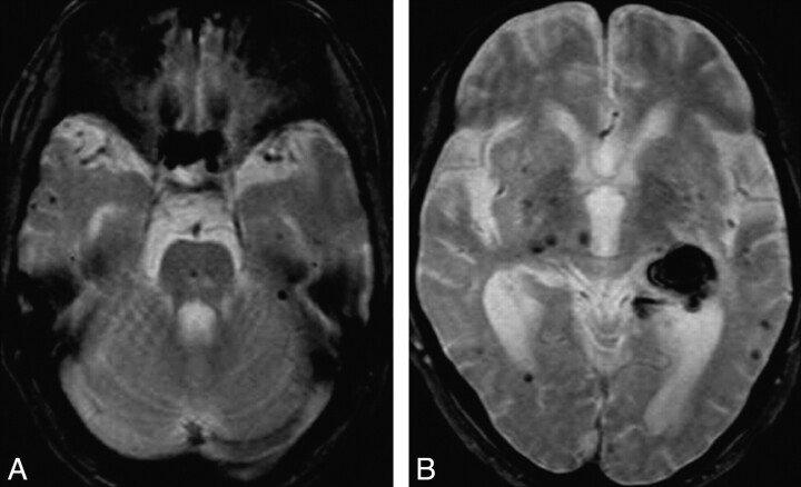
Background and purpose: Although accumulating evidence suggests the presence of microbleeds as a risk factor for intracerebral hemorrhage (ICH), little is known about its significance in anticoagulated patients. The aim of this study was to determine whether the presence of microbleeds is associated with recurrent hemorrhagic stroke in patients who had received warfarin following atrial fibrillation-associated cardioembolic infarction.
Materials and methods: A total of 87 consecutive patients with acute recurrent stroke, including 15 patients with ICH and 72 patients with cerebral infarction, were enrolled in this study. International normalized ratios (INRs), vascular risk factors, and imaging characteristics, including microbleeds on T2*-weighted MR images and white matter hyperintensity (WMH) on T2-weighted MR images, were compared in the 2 groups.
Results: Microbleeds were noted more frequently in patients with ICH than in patients with cerebral infarction (86.7% versus 38.9%, P = .0007). The number of microbleeds was larger in patients with ICH than in patients with cerebral infarction (mean, 8.4 versus 2.1; P = .0001). INR was higher in patients with ICH than in patients with cerebral infarction (mean, 2.2 versus 1.4; P < .0001). The frequency of hypertension was higher in patients with ICH than in patients with cerebral infarction (86.7% versus 45.8%, P = .0039). Multivariate analysis revealed that the presence of cerebral microbleeds (odds ratio, 7.383; 95% confidence interval, 1.052-51.830) was associated with ICH independent of increased INR and hypertension.
Conclusion: The presence of cerebral microbleeds may be an independent risk factor for warfarin-related ICH, but more study is needed because of strong confounding associations with elevated INR and hypertension.
Figures

References
-
- Tanaka A, Ueno Y, Nakayama Y, et al. Small chronic hemorrhages and ischemic lesions in association with spontaneous intracerebral hematomas. Stroke 1999;30:1637–42 - PubMed
-
- Greenberg SM, O'Donnell HC, Schaefer PW, et al. MRI detection of new hemorrhages: potential marker of progression in cerebral amyloid angiopathy. Neurology 1999;53:1135–38 - PubMed
-
- Roob G, Lechner A, Schmidt R, et al. Frequency and location of microbleeds in patients with primary intracerebral hemorrhage. Stroke 2000;31:2665–69 - PubMed
-
- Kinoshita T, Okudera T, Tamura H, et al. Assessment of lacunar hemorrhage associated with hypertensive stroke by echo-planar gradient-echo T2*-weighted MRI. Stroke 2000;31:1646–50 - PubMed
Publication types
MeSH terms
Substances
LinkOut - more resources
Full Text Sources
Medical