. 2008 Aug;26(8):2042-51.
doi: 10.1634/stemcells.2008-0149. Epub 2008 May 22.

Hematopoietic stem cells regulate mesenchymal stromal cell induction into osteoblasts thereby participating in the formation of the stem cell niche

Affiliations

Hematopoietic stem cells regulate mesenchymal stromal cell induction into osteoblasts thereby participating in the formation of the stem cell niche

Younghun Jung et al. Stem Cells. 2008 Aug.

Abstract

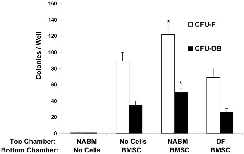
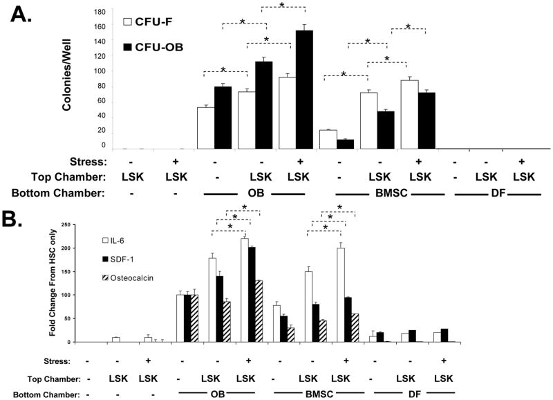
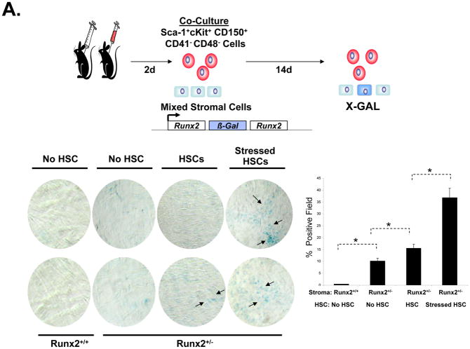
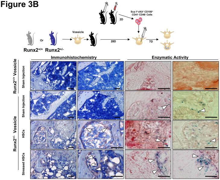
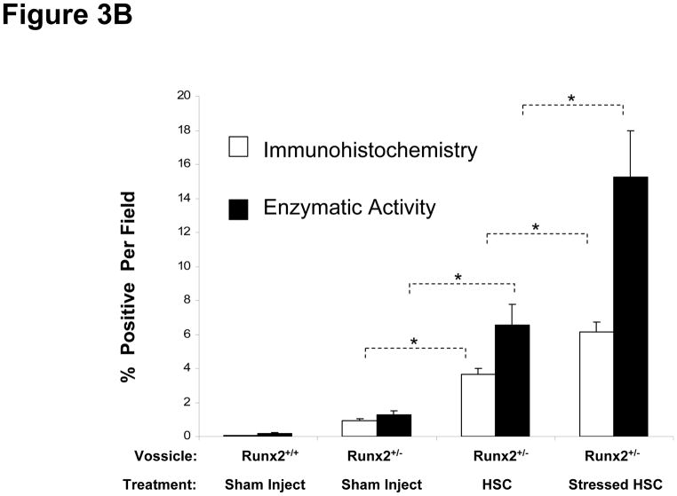
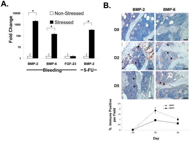
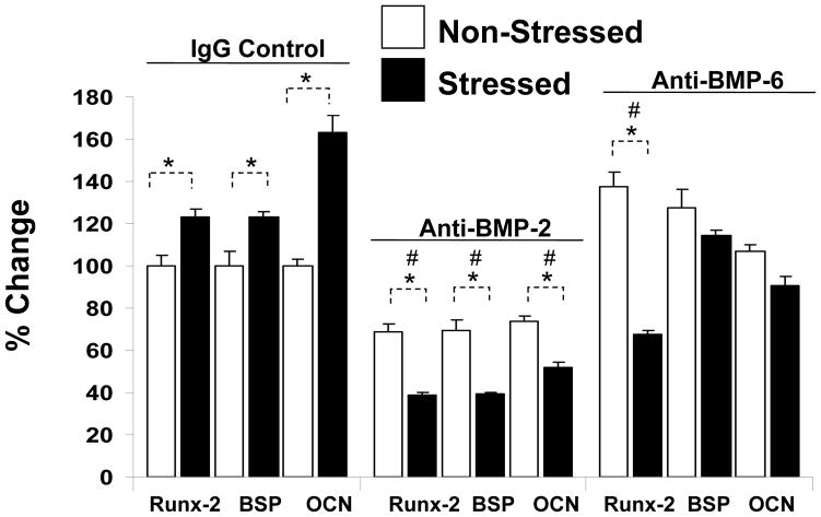
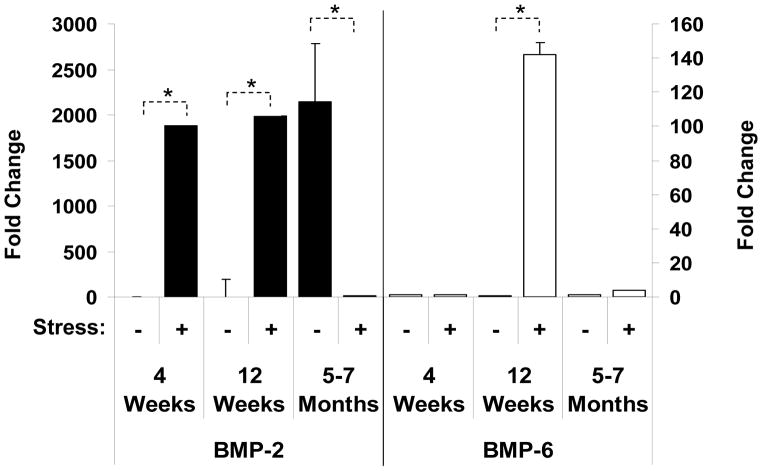
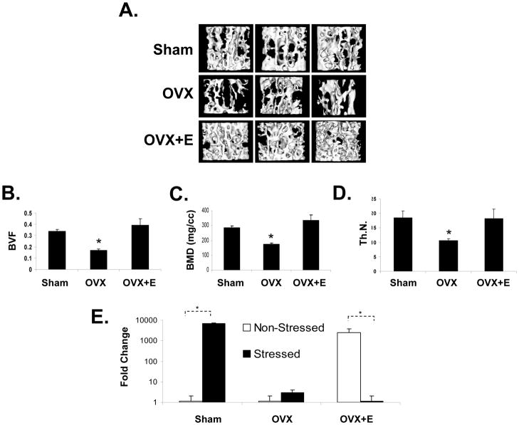
Crosstalk between hematopoietic stem cells (HSCs) and the cells comprising the niche is critical for maintaining stem cell activities. Yet little evidence supports the concept that HSCs regulate development of the niche. Here, the ability of HSCs to directly regulate endosteal development was examined. Marrow was isolated 48 hours after "stressing" mice with a single acute bleed or from control nonstressed animals. "Stressed" and "nonstressed" HSCs were cocultured with bone marrow stromal cells to map mesenchymal fate. The data suggest that HSCs are able to guide mesenchymal differentiation toward the osteoblastic lineage under basal conditions. HSCs isolated from animals subjected to an acute stress were significantly better at inducing osteoblastic differentiation in vitro and in vivo than those from control animals. Importantly, HSC-derived bone morphogenic protein 2 (BMP-2) and BMP-6 were responsible for these activities. Furthermore, significant differences in the ability of HSCs to generate a BMP response following stress were noted in aged and in osteoporotic animals. Together these data suggest a coupling between HSC functions and bone turnover as in aging and in osteoporosis. For the first time, these results demonstrate that HSCs do not rest passively in their niche. Instead, they directly participate in bone formation and niche activities. Disclosure of potential conflicts of interest is found at the end of this article.


Conflict of interest statement

Competing Interests Statement

The authors declare that they have no competing financial interests.

Figures

Figure 1
Figure 1. Non-Adherent Bone Marrow Fractions Alter CFU-F/CFU-OB Formation
Non-adherent bone marrow cell fractions (NABM) were evaluated for their effects on the generation of fibroblastic (CFU-F) and osteoblastic (CFU-OB) colonies from BMSCs in vitro. Cells from primary bone marrow stromal cell (BMSC) cultures were trypsinized, resuspended and replated at 100 cells/well. NABM cells were collected from marrow and plated on plastic for 24 hours, recovered and washed, and placed into the top chamber of a chambered culture plate to exclude the possibility that cells from the non-adherent bone marrow fraction may contribute to colony formation. Newborn dermal fibroblasts (DF) were placed into the top chamber of the culture to served as negative controls. Cultures of just the NABM were also included. After 21 days the colonies (>50 cells) were quantified as either CFU-F or CFU-OB using methyl violet or Von Kossa staining. The data are presented as mean ± s.d. for n=6 determinations in three independent investigations. *p <0.05 compared to no cells in the top well (Students t-test).
Figure 2
Figure 2. Hematopoietic Stress Induces Cytokine and Mesenchymal Changes in the HSC niche
In (A) LinSca-1+cKit+ (LSK) cells were isolated from hematopoetically ‘stressed’ (removing ~20–30% of the calculated blood volume by jugular vein venipucture) (+) and ‘non-stressed’ (puncture only) (−) at 48 hour and were added to murine osteoblasts (OBs), bone marrow stromal cells (BMSC) or dermal fibroblasts (DF) in dual chambered culture plates. At 21 days, the cultures were examined for fibroblastic (CFU-F, methylviolet staining) or osteoblastic (CFU-OBs, Von Kossa staining) colonies. In (B) the CM was assayed for IL-6, SDF-1 by ELISA (R&D Systems), or the bone specific protein osteocalcin by RIA (Biomedical Technologies, Inc. Stoughton, MA), and are expressed as % change from OB levels alone for each assay,where *p <0.05 (ANOVA). The data indicate that HSCs from hematopoietically stressed animals are able to direct the formation of a microenvironment directly and influence cytokine secretion of stroma itself.
Figure 3
Figure 3. Sca-1+cKit+CD150+CD41CD48 Bone Marrow Cells Regulate Stromal Cell Fate In Vitro and In Vivo
In (A) Sca-1+cKit+CD150+CD41CD48 bone marrow cells were isolated from hematopoetically ‘stressed’ and ‘non-stressed’ (puncture only) (−) animals at 48 hours and were directly added to pre-established murine BMSC derived from Runx2 knock-in animals. FACs profiles of the sorted HSCs are presented in Supplemental Figure 1. After 14 days, β-galactosidase activity was detected. The data are presented as mean ± s.d. for n=5 determinations in three independent investigations. In (B), vossicles derived from Runx2 knock-in animals or littermate controls were implanted into wild-type animals. Four vossicles (n=4) were in of each five mice (n=5). At one month, the vossicles were exposed and either injected with 500 Sca-1+cKit+CD150+CD41CD48 bone marrow cells isolated from hematopoetically stressed or ‘non-stressed’ animals, or sham injected with PBS. One week later, the vossicles were harvested and tissues stained for β-galactosidase by immunohistochemistry or for enzymatic activity (Bottom Left). Bar=50 microns. IgG control stained tissues are not presented but did not differ from sham injected knock-in tissues. Quantification of the data is presented in the Bottom Right panel. The data demonstrate that Sca-1+cKit+CD150+CD41CD48 induce Runx2 expression in vitro and in vivo.
Figure 4
Figure 4. Hematopoietic Stress Induces BMP-2 and BMP-6 Expression in the Marrow and HSCs
In (A) real-time RT-PCR was used to evaluate mRNA levels for BMP-2, BMP-6 and FGF-23 from HSCs (Sca-1+cKit+CD150+CD41CD48 cells) isolated 2 days following an acute stress (jugular vein puncture and aspiration, versus puncture only, or 5-FU versus vehicle). The cells were lysed and RNA prepared for evaluation of mRNA levels by reai-time RT-PCR. The data is presented as fold change normalized to GAPDH, where the expression level derived from the non-stressed animals was set as the standard. *p < 0.05 versus the non-stressed animals (Krusal-Wallis test, and Dunn’s multiple comparisons test). In (B) bones were harvested at 0, 2 and 5 days following an acute stress, fixed, decalcified and stained with an antibody to BMP-2 and BMP-6 or an IgG control (not shown) in conjunction with a HRP-AEC staining system and counter stained with hematoxylin and eosin. Bar=100 microns. The data demonstrate that BMP-2 and BMP-6 are expressed by HSCs and the marrow following stress. Quantification of the data is presented in the Bottom Right panel.
Figure 5
Figure 5. HSCs Regulate Mesenchymal Fate Through BMPs
Co-culture investigations were established by placing HSCs (Sca-1+cKit+CD150+CD41CD48) derived from stressed or non-stressed animals in the top chamber of a dual culture plate, and mixed BMSCs in the bottom well, in the presence or absence of neutralizing antibody to BMP-2 or BMP-6, or an IgG isotype matched control each added daily at 5 ng/ml. After 21 days the OB phenotype was examined by real-time RT-PCR for the expression of the Runx2 (an OB specific transcription factor), or the OB specific proteins bone sialoprotein (BSP) or osteocalcin (OCN). The data is presented as % change normalized to GAPDH, where the expression levels of co-cultures of BMSCs/HSCs (from the non-stressed animals) was set as the standard. *p <0.05 between HSCs derived from stressed and non-stressed animals, and # signifies differences between anti-BMP and IgG treated controls (p<0.05, Krusal-Wallis test, and Dunn’s multiple comparisons test). The data demonstrate that blockade of BMP-2 or BMP-6 activities modifies lineage differentiation.
Figure 6
Figure 6. Aging Alters the BMP Response in HSCs
HSCs (Sca-1+cKit+CD150+CD41CD48) were isolated 2 days following an acute stress from young (4 weeks), sexually mature (12 weeks) and aged (5–7 months) mice. The cells were lysed and RNA prepared for evaluation by real-time RT- PCR. The data are presented as fold change normalized to GAPDH, where the expression levels of the HSCs derived from the non-stressed animals was set as the standard. *p < 0.05 versus the non-stressed animals.
Figure 7
Figure 7. Ovarectomy Alters the BMP response in HSCs
To explore the possibility that bone loss in osteoporosis may be a consequence of defective HSC function, mice were either sham operated (n=10), ovariectomized (OVX, n=10), or treated with OVX with hormone replacement (β-estradiol or E, n=10). At two months, (A) 3-dimensional micro-CT measurements of the first lumbar vertebrate were performed. (B) Bone volume fraction (BVF), (C) bone mineral density (BMD), (D) trabecular number (Th.N.) were also significantly reduced in the OVX treated animals compared to controls or OVX + E treated groups. (E) HSCs derived from sham treated animals responded to a hematopoietic stress by generating a BMP-2 response. The data are presented as fold change normalized to GAPDH, where the expression level of derived from the non-stressed animals was set as the standard. * *p < 0.05 versus the non-stressed animals (Krusal-Wallis test, and Dunn’s multiple comparisons test).

Similar articles

Cited by

References

    1. Taichman RS, Emerson SG. Human osteoblasts support hematopoiesis through the production of granulocyte colony-stimulating factor. J Exp Med. 1994;179:1677–1682. - PMC - PubMed
    1. Adams GB, Chabner KT, Alley IR, Olson DP, Szczepiorkowski ZM, Poznansky MC, Kos CH, Pollak MR, Brown EM, Scadden DT. Stem cell engraftment at the endosteal niche is specified by the calcium-sensing receptor. Nature. 2006;439(7076):599–603. - PubMed
    1. Arai F, Hirao A, Ohmura M, Sato H, Matsuoka S, Takubo K, Ito K, Koh GY, Suda T. Tie2/Angiopoietin-1 Signaling Regulates Hematopoietic Stem Cell Quiescence in the Bone Marrow Niche. Cell. 2004;118(2):149–161. - PubMed
    1. Calvi LM, Adams GB, Weibrecht KW, Weber JM, Olson DP, Knight MC, Martin RP, Schipani E, Divieti P, Bringhurst FR, Milner LA, Kronenberg HM, Scadden DT. Osteoblastic cells regulate the haematopoietic stem cell niche. Nature. 2003;425(6960):841–846. - PubMed
    1. Nilsson SK, Johnston HM, Whitty GA, Williams B, Webb RJ, Denhardt DT, Bertoncello I, Bendall LJ, Simmons PJ, Haylock DN. Osteopontin, a key component of the hematopoietic stem cell niche and regulator of primitive hematopoietic progenitor cells. Blood. 2005:2004–2011. - PubMed

Publication types

Substances