. 2008 Jul;16(7):1208-16.
doi: 10.1038/mt.2008.104. Epub 2008 May 20.

MicroRNAs regulate ocular neovascularization

Affiliations

MicroRNAs regulate ocular neovascularization

Jikui Shen et al. Mol Ther. 2008 Jul.

Abstract

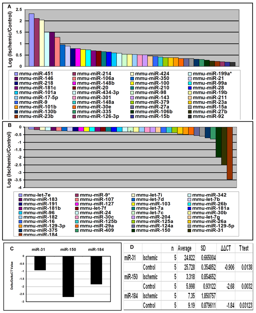
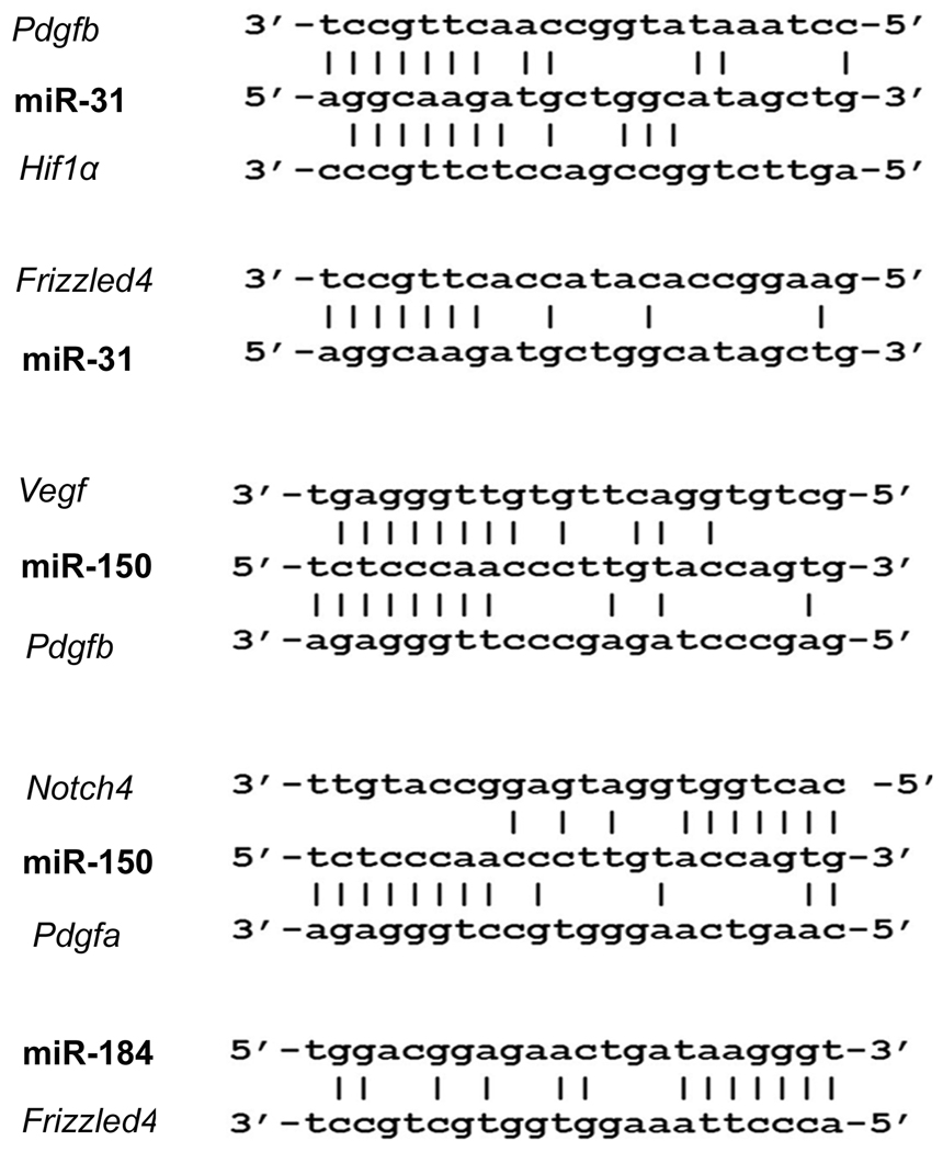
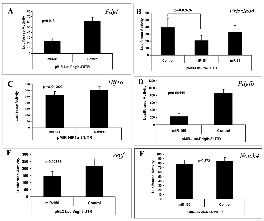
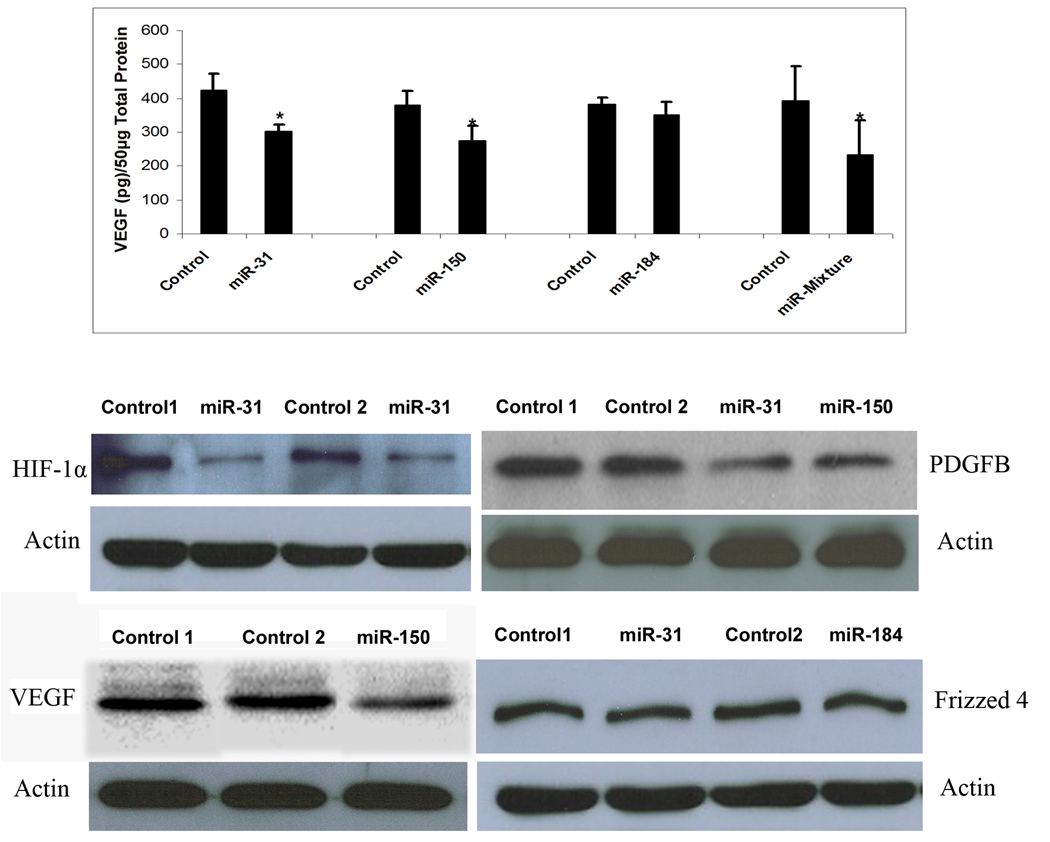
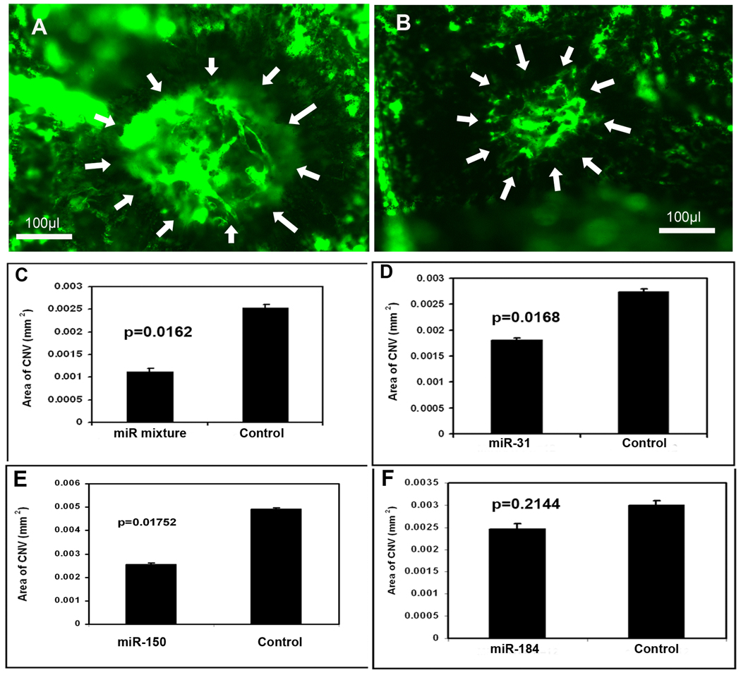
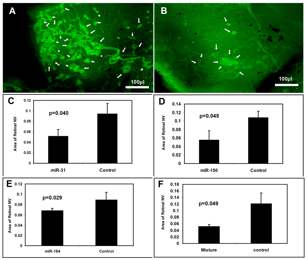
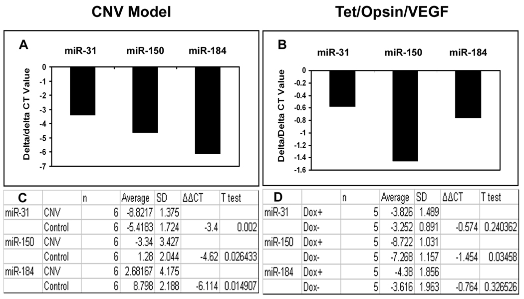
In this study, we used ischemia-induced retinal neovascularization (NV) as a model to investigate the possible role of microRNAs in a clinically important disease process. Microarray analysis demonstrated seven microRNAs (miR-106a, -146, -181, -199a, -214, -424, and -451) that were substantially increased and three microRNAs (miR-31, -150, and -184) that were substantially decreased in ischemic retina. Potential targets for the upregulated microRNAs were not identified, but bioinformatic analysis suggested target genes for the downregulated microRNAs, and these were confirmed using a luciferase reporter assay. Real-time reverse transcriptase PCR confirmed that the substantial levels of miR-31, -150, and -184 present in normal retina were significantly reduced in ischemic retina. Interestingly, constitutive levels of miR-31 and -184 are high in the cornea and lens, two avascular tissues. Intraocular injection of pre-miR-31, -150, or -184 significantly reduced ischemia-induced retinal NV, and injection of pre-miR-31 or -150 also significantly reduced choroidal NV. These data suggest that alteration of microRNA levels contributes to two types of ocular NV, and that injection or enhanced expression of microRNAs is a potential therapeutic strategy.


Figures

Figure 1
Figure 1. Identification of microRNAs that are upregulated or downregulated in ischemic retina
Six C57BL/6 mice were placed in 75% oxygen at P7 and returned to room air at P12. At P15, mice were euthanized and small RNA species were isolated from the retinas. The RNA samples from ischemic retinas were pooled and labeled with Cy3 and the samples from control retinas were pooled and labeled with Cy5. The RNA probes were hybridized with microarrays as described in Methods. The log2 of the ratio of Cy3/Cy5 signal strength is shown for 40 microRNAs that were upregulated in ischemic retina (A) and 37 were that were downregulated (B). Real time RT-PCR was used to independently determine levels of miR-31, miR-150, and miR-184 relative to levels of 5s rRNA. The mean threshold cycle number (delta CT) for miR-31, miR-150, and miR-184 was determined for RNA from ischemic and control retinas (n=5 for each) and the mean difference (delta/delta CT) ranged from about −1.0 (miR-31) to −2.5 (miR-150, C). Statistical comparisons confirmed that each of the miRs were significantly reduced in ischemic compared to control retinas (D).
Figure 2
Figure 2. Identification of target genes for down-regulated microRNAs
The European DNA database was searched for 7–8 consecutive complementary sequences starting 2 bp from the 5’ or 3’ end of miR-31, miR-184, and miR-150. Complementary regions were identified in the 3’UTR of: Pdgfb, Hif1α, and Frizzled4 for miR-31; Vegf, Pdgfb, Pdgfa, and Notch4 for miR-150, and Frizzled 4 for miR-184 (A). Areas of alignment with the 3’UTR of Vegf, Pdfgb, Hif1a and Notch4, but not Frizzled4 were highly conserved among different species.
Figure 3
Figure 3. The effect of microRNAs on promoter activity of purported target genes
Luciferase reporter constructs were generated for microRNA reporter assays for each of the potential target sequences listed in Figure 2 except Pdgfa. Each bar represents the mean (±SEM) calculated from three experiments. Compared to a control microRNA with scrambled sequence, miR-31 significantly reduced luciferase expression for pMIR-Luc-Pdgfb-3’UTR (A) and pMIR-Luc-HIF1α-3’UTR (B), but not pMIR-Luc-Fz4-3’UTR (C). Luciferase expression was significantly reduced by miR-184 for pMIR-Luc-Fz4-3’UTR (C). Luciferase expression was reduced by miR-150 for pMIR-Luc-Pdgfb-3’UTR (D) and pGL2-Luc-Vegf-3’UTR (E), but not pMIR-Luc-Notch4-3’UTR (F). Statistical comparisons were made by paired t-test.
Figure 4
Figure 4. Effect of microRNAs on target gene product levels in ischemic retina
C57BL/6 mice were placed in 75% oxygen at P7 and returned to room air at P12 and given an intravitreous injection of 1 µl of 5 µM experimental precursor microRNA (pre-microRNA) in one eye and 1 µl of 5 µM control pre-microRNA in the other eye. At P15, mice were euthanized and VEGF was measured in retinal homogenates by ELISA (top panel). Each bar represents the mean (±SEM) calculated from 3 experiments. VEGF protein was not detectable in retinas from mice in room air at P15 (n=7), but high levels were measured in ischemic retinas (n=10). Compared to injection of control pre-microRNA, injection of pre-miR-31 or -150, but not pre-miR-184 caused significant reduction in VEGF in ischemic retina (top panel). There was also a significant reduction in VEGF in ischemic retinas from eyes injected with 1 µl containing 1.67 µM concentrations of each of the 3 experimental pre-microRNAs compared to fellow eyes injected with 1 µl containing 5 µM control pre-microRNA. Immunoblots of retinal homogenates from eyes injected with pre-miR-31 showed reductions in HIF1α (middle panel, left) and PDGF-B (middle panel, right) compared to eyes injected with control pre-microRNA. Ischemic retinas from eyes injected with pre-miR-150 showed reductions in PDGF-B (middle panel, right) and VEGF (bottom panel, left). Frizzled 4 was not reduced by injection of pre-miR-31 or -184 (bottom panel, right). Each immunoblot was repeated at least once with similar results.
Figure 5
Figure 5. Effect of microRNAs on ischemia-induced retinal neovascularization (NV)
At P7, litters of C57BL/6 mice were placed in 75% oxygen. At P12, mice (n=8) were given an intraocular injection of experimental precursor microRNA (pre-microRNA) or a mixture of all 3 experimental pre-microRNAs in the right eye and control pre-microRNA in the other eye. At P17, retinal NV was stained as described in Methods and measured on retinal flat mounts by image analysis. Compared to the retinas from mice injected with control pre-microRNA, which showed prominent NV on the surface of the retina (A, arrows), retinas from mice injected with a mixture of pre-miR-31, -150, and -184 showed substantially less NV (B, arrows). Measurements done by image analysis showed that the mean area of NV was significantly less for eyes injected with pre-miR-31 (C), -150 (D), -184 (E), or a mixture of all three (F) compared to fellow eyes injected with control pre-microRNA. Each bar represents the mean (±SD) area of NV calculated from 8 mice. Statistical comparisons were made by paired t-test.
Figure 6
Figure 6. Effect of microRNAs on choroidal neovascularization (NV)
Adult C57BL/6 mice had rupture of Bruch’s membrane by laser photocoagulation in three locations in each eye and they were given an intraocular injection of 1 µl of 5 µM precursor microRNA (pre-microRNA) in one eye and 1 µl of control pre-microRNA in the fellow eye on day 0 and day 7. On day 14, mice were perfused with fluorescein-labeled dextran and the area of choroidal NV at Bruch’s membrane rupture sites was measured by image analysis. Each bar represents the mean (±SD) area of choroidal NV calculated from at least 12 experimental values. Compared to the area of NV in eyes injected with control pre-microRNA (A), there was a significant decrease in eyes injected with a mixture of all 3 experimental pre-microRNAs (B and C), pre-miR-31 (D), or -150 (E), but not pre-miR-184 (F). Statistical comparisons were made by paired t-test.
Figure 7
Figure 7. Expression of miR-31, miR-150 and miR-184 in other models of ocular neovascularization
C57BL/6 mice (n=6) had laser-induced rupture of Bruch’s membrane at 10 locations in one eye and after 4 days were euthanized, anterior segments were removed from each eye, and small RNAs were isolated from eye cups. Double transgenic Tet/opsin/VEGF mice with doxycycline-inducible expression of VEGF in the retina were given 2 mg/ml of doxycycline in their drinking water (n=5) or maintained on normal water (n=5, controls). After 3 days, the mice were euthanized and small RNAs were isolated from the retinas. Real time RT-PCR for miR-31, -150, -184 and 5S rRNA was done as described in Methods. The mean threshold cycle number (delta CT) for miR-31, miR-150, and miR-184 was determined for RNA isolated from experimental and control eyes and the bars show the mean difference (delta/delta CT). Compared to contralateral control eyes, those with choroidal NV showed substantial reductions in mean threshold number for each of the miRs (A) and statistical comparisons confirmed that the levels of miR-31, -150, and -184 were all significantly reduced in eyes with choroidal NV (C). Compared to untreated Tet/opsin/VEGF mice, those treated with doxycycline resulting in expression of VEGF in the retina showed minimal changes in mean threshold number for miR-31 and -184 that were not statistically significant, but there was a significant reduction in the level of miR-150 (B and D).

References

    1. Campochiaro PA. Ocular versus extraocular neovascularization: mirror images or vague resemblances. Invest. Ophthalmol. Vis. Sci. 2006;47:462–474. - PubMed
    1. Campochiaro PA. Ocular neovascularisation and excessive vascular permeability. Expert. Opin. Biol. Ther. 2004;4:1395–1402. - PubMed
    1. Aiello LP, Pierce EA, Foley ED, et al. Suppression of retinal neovascularization in vivo by inhibition of vascular endothelial growth factor (VEGF) using soluble VEGF-receptor chimeric proteins. Proc. Natl. Acad. Sci. U.S.A. 1995;92:10457–10461. - PMC - PubMed
    1. Miller JW, Adamis AP, Shima DT, et al. Vascular endothelial growth factor/vascular permeability factor is temporally and spatially correlated with ocular angiogenesis in a primate model. Am. J. Pathol. 1994;145:574–584. - PMC - PubMed
    1. Ozaki H, Seo M-S, Ozaki K, et al. Blockade of vascular endothelial cell growth factor receptor signaling is sufficient to completely prevent retinal neovascularization. Am. J. Pathol. 2000;156:679–707. - PMC - PubMed

Publication types

LinkOut - more resources