. 2008 Jun;26(6):695-701.
doi: 10.1038/nbt1398. Epub 2008 May 25.

Targeted gene inactivation in zebrafish using engineered zinc-finger nucleases

Affiliations

Targeted gene inactivation in zebrafish using engineered zinc-finger nucleases

Xiangdong Meng et al. Nat Biotechnol. 2008 Jun.

Abstract

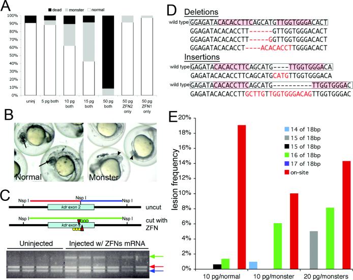
Direct genomic manipulation at a specific locus is still not feasible in most vertebrate model organisms. In vertebrate cell lines, genomic lesions at a specific site have been introduced using zinc-finger nucleases (ZFNs). Here we adapt this technology to create targeted mutations in the zebrafish germ line. ZFNs were engineered that recognize sequences in the zebrafish ortholog of the vascular endothelial growth factor-2 receptor, kdr (also known as kdra). Co-injection of mRNAs encoding these ZFNs into one-cell-stage zebrafish embryos led to mutagenic lesions at the target site that were transmitted through the germ line with high frequency. The use of engineered ZFNs to introduce heritable mutations into a genome obviates the need for embryonic stem cell lines and should be applicable to most animal species for which early-stage embryos are easily accessible.


Figures

Figure 1
Figure 1
Engineering ZFNs that target kdra exon 2 using a bacterial one-hybrid system. a. First selection stage to identify single zinc fingers with optimized binding to each 3-bp subsite within a recognition element. b. Recombination of individual fingers and subsequent second selection stage against full ZFP recognition elements. c. Sequence logos are shown for the binding specificity of ZFPs incorporated into ZFNs for subsequent analysis in zebrafish embryos. d. Overview of approach for ZFN-targeted mutagenesis in zebrafish embryos.
Figure 2
Figure 2
Effects of ZFN mRNA injections on zebrafish embryos. a. Graph depicting proportion of embryos with indicated phenotypes associated with increasing doses of injected ZFN mRNA. b. Examples of normal (left panel) and abnormal (“monster”, right panel) embryos injected with 15 pg of each ZFN mRNA. Left panel, arrows indicate position of the eye. Right panel, arrowhead indicates defective eye formation and severe necrosis. c. PCR genotyping to assay presence or absence of NspI site. Wild type fragments indicative of the presence of the exon 2 NspI site are indicated by red and blue arrows. The larger undigested fragment caused by loss of the NspI site is indicated by a green arrow. d. Lesions observed in morphologically normal embryos injected with 15 pg of both ZFN mRNAs. Boxes indicate wild type kdra sequence. Pink shaded boxes label the ZFN recognition elements. Red dashes or letters indicate the positions of deletions or insertions, respectively. e. Graph depicting lesion frequency at the kdra exon 2 site and a subset of off-site targets in ZFN-injected embryos. Lesion frequency is proportion of sequences containing indels at the target over total number of sequences for a given site. Data for each group of embryos were obtained using Solexa sequencing as described in Materials and Methods.
Figure 3
Figure 3
Segmental artery defects in ZFN-mutation bearing embryos. a., b. Phenotypically wild type embryo from a cross between founder 883.2M and a female heterozygous for kdraum6. b. Normal segmental artery formation; segmental arteries are indicated by white arrows. c., d. Sibling of embryo in a. displaying normal morphology, but a failure to form segmental arteries. e., f. Normal morphology but partial segmental artery formation (white arrows) in a phenotypically mutant embryo from a cross between founder 889.3M and a female heterozygous for kdraum6. g.,h. Mutant embryo from an incross of kdraum6 heterozygous parents. h. Loss of segmental arteries in a kdraum6 mutant embryo. b, d, f, h – confocal images of embryos bearing the Tg(fli1:egfp)y1 transgene.
Figure 4
Figure 4
Genotypic characterization of ZFN-induced mutations. a. NspI PCR genotyping assay on individual embryos derived from founders 889.7M and 889.1M. Embryos were genotyped for kdraum6 by sequencing (see Supplementary Fig. 10 for chromatograms); +/+ indicates homozygous wild type and +/− heterozygous for the um6 allele; M – male, F – female. b. CelI nuclease genotyping of individual embryos derived from founder 889.1M; embryos are the same as those shown in a. c. Genetic lesions identified in mutant embryos derived from individual crosses between indicated founder and a kdraum6 heterozygote. ZFN recognition elements are shaded in pink and the ZFN cleavage position on this strand is indicated by an arrow. Gray dashes or letters indicate the positions of deletions or insertions, respectively, in the progeny sequences.

Comment in

  • Targeted mutagenesis in zebrafish.
    Woods IG, Schier AF. Woods IG, et al. Nat Biotechnol. 2008 Jun;26(6):650-1. doi: 10.1038/nbt0608-650. Nat Biotechnol. 2008. PMID: 18536686 Free PMC article. No abstract available.

References

    1. Porteus MH, Carroll D. Gene targeting using zinc finger nucleases. Nat Biotechnol. 2005;23:967–973. - PubMed
    1. Urnov FD, et al. Highly efficient endogenous human gene correction using designed zinc-finger nucleases. Nature. 2005;435:646–651. - PubMed
    1. Porteus MH, Baltimore D. Chimeric nucleases stimulate gene targeting in human cells. Science. 2003;300:763. - PubMed
    1. Koller BH, Smithies O. Inactivating the beta 2-microglobulin locus in mouse embryonic stem cells by homologous recombination. Proceedings of the National Academy of Sciences of the United States of America. 1989;86:8932–8935. - PMC - PubMed
    1. Zijlstra M, Li E, Sajjadi F, Subramani S, Jaenisch R. Germ-line transmission of a disrupted beta 2-microglobulin gene produced by homologous recombination in embryonic stem cells. Nature. 1989;342:435–438. - PubMed

Publication types

MeSH terms