. 2008 Jun;15(6):634-40.
doi: 10.1038/nsmb.1420. Epub 2008 May 25.

Triple-helix structure in telomerase RNA contributes to catalysis

Affiliations

Triple-helix structure in telomerase RNA contributes to catalysis

Feng Qiao et al. Nat Struct Mol Biol. 2008 Jun.

Abstract

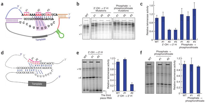
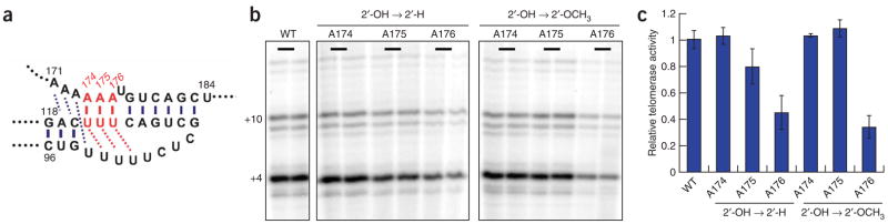
Telomerase is responsible for replication of the ends of linear chromosomes in most eukaryotes. Its intrinsic RNA subunit provides the template for synthesis of telomeric DNA by the reverse-transcriptase (TERT) subunit and tethers other proteins into the ribonucleoprotein (RNP) complex. We report that a phylogenetically conserved triple helix within a pseudoknot structure of this RNA contributes to telomerase activity but not by binding the TERT protein. Instead, 2'-OH groups protruding from the triple helix participate in both yeast and human telomerase catalysis; they may orient the primer-template relative to the active site in a manner analogous to group I ribozymes. The role of RNA in telomerase catalysis may have been acquired relatively recently or, alternatively, telomerase may be a molecular fossil representing an evolutionary link between RNA enzymes and RNP enzymes.


Figures

Figure 1
Figure 1
The core region of the S. cerevisiae telomerase RNA has a conserved pseudoknot RNA structure. (a) One of the four secondary-structure models (see Supplementary Fig. 1 for all four models) of the S. cerevisiae telomerase RNA core region. Numbers 1 to 12 refer to twelve scanning mutants made to test the four models. In each of these mutants, eight consecutive nucleotides were mutated to their Watson-Crick complementary bases. The 5′ and 3′ end nucleotides are labeled according to the numbering system in ref. . (b) In vitro telomerase activities of the 12 scanning mutants. A schematic illustration of the alignment of telomeric DNA substrate with the template region of the telomerase RNA is shown above. Numbers at the bottom of the gel denote the relative activity of each mutant calculated based on the two independent reactions shown. The +1 marker was the 15-nt single-stranded telomeric primer extended by 1 nt with the addition of [33P]ddTTP and terminal deoxynucleotidyltransferase. WT, wild type. (c) Compensatory mutational analysis confirms Stem 1 and Stem 2 formation in the pseudoknot region of the S. cerevisiae telomerase RNA. In vitro telomerase activities of the Stem 1–disrupting mutants 2a (723GGUUU728→CCAAA), 2b (729AUUCU733→UAAGA), 8a (771AGAAU775→UCUUA), 8b (776AAAUCC781→UUUAGG) and the Stem 1–restoring mutants 8b/2a (combination of mutants 8b and 2a) and 8a/2b (combine mutants 8a and 2b). In vitro telomerase activities of the Stem 2–disrupting mutants 5a (751AGUAGA756→UCAUCU) and 12a (807UCUAUU812→AGAUGA) and the Stem 2–restoring mutant 12a/5a (combination of mutants 12a and 5a). The activities of individual mutants were normalized to the wild-type activity. Nucleotides are labeled according to the numbering system in ref. .
Figure 2
Figure 2
The pseudoknot region of the S. cerevisiae telomerase RNA contains a triple-helix structure. (a) The sequences and the secondary structures of the triple-helix and adjacent regions in the context of the Micro-T 170 telomerase RNA (see Supplementary Fig. 3 for detail). The three dotted lines indicate the base triples best supported by this work; an additional flanking U-A•U triple may also form but was not tested here. (b) Base triples formed by U-A•U, C-G•C and C-G•U, respectively. All involve formation of Hoogsteen base pairs in the major groove of the Watson-Crick duplex. In the case of the C-G•C triple, protonation of N3 of cytosine at pH below its pKa (free nucleotide has pKa ~4.2) could provide an additional hydrogen bond (gray). (c) In vitro telomerase activities of the telomerase RNA triple helix–disrupting mutants 11c_3G (A804–A806→GGG), 6a_3C (U757–A759→CCC), 4b_3C (U746–U748→CCC) and the triple helix–restoring mutants 3GCC-1 (A804–A806→GGG/U757–A759→CCC/U746–U748→CCC), 3GCC-2 (A804–A806→GGG/U757–A759→CCC/U745–U747→CCC) and 3GCC-3 (A804–A806→GGG/U757–A759→CCC/U744–U746→CCC). The activities of individual mutants were normalized to the wild-type (WT) activity. (d) Telomere-length analysis of the telomerase RNA triple helix–disrupting mutants and triple helix–restoring mutants compared with the wild-type TLC1. Southern blot shows lengths of telomeric XhoI restriction fragments from cells harboring different TLC1 constructs and grown for 125 generations. Duplicate lanes represent two independent clones for each mutant. A probe that hybridizes to both strands of telomeric DNA was used. The markers shown were radiolabeled with 2-Log DNA Ladder (NEB). Y′ telomeres are preceded by a repeated DNA sequence that contains an XhoI restriction site.
Figure 3
Figure 3
The triple-helix structure in TLC1 is not responsible for the binding affinity of the reverse-transcriptase subunit, Est2. ProA-Est2 translated in rabbit reticulocyte lysate (RRL) was assembled with wild-type (WT) Micro-T 170 telomerase RNA and various mutants shown in Figs. 1c and 2a. The assembled telomerase RNP was immunoprecipitated, and the associated telomerase RNA was separated by denaturing PAGE. The amounts of the Est2-bound RNA (b) and the input RNA (c) were quantified from the band intensity using a PhosphorImager and ImageQuant TL. The relative efficiency of Est2 binding is calculated from the ratio of the bound RNA to the input RNA compared to WT. P4P6 RNA (158 nt), an autonomously folded domain of group I intron ribozyme, was used as a negative control for Est2 binding. Error bars, s.d.
Figure 4
Figure 4
The triple-helix region is in close proximity to the 3′ end of the DNA substrate. (a) The secondary-structure model of the telomerase RNA Micro-T is shown with an annealed DNA primer—the substrate for telomerase—coupled through a thiophosphate to a photoactivatable cross-linking azidophenacyl group. Upon irradiation at 302 nm, the azido group is converted to a nitrene, which reacts to form a covalent bond between the DNA primer and nearby regions of the RNA. (b) Cross-linked and free Micro-T RNAs separated by denaturing PAGE. A primer without the azidophenacyl label gave no cross-link after UV irradiation, nor did Micro-T alone. (c,d) Primer extension analyses of DNA–Micro-T RNA conjugates with a 5′–32P-labeled oligonucleotide complementary to the 3′-terminal sequences of the Micro-T RNA. Lanes U, A, C and G correspond to sequencing reactions with non–cross-linked RNA template. The other lanes show primer extension reactions using various cross-linked or irradiated but non–cross-linked RNA species, as indicated. The major termination sites of primer extension, at which reverse transcriptase pauses or terminates because of the cross-linked nucleotides, are boxed in green in c and red in d, and the corresponding positions on the secondary-structure model are indicated by green or red arrowheads in a. Nucleotides are labeled according to the numbering system in ref. .
Figure 5
Figure 5
The 2′-OH groups in the triple-helix regions of both yeast and human telomerase RNAs are important for catalysis. (ac) Both 2′-OH to 2′-H and phosphate to phosphorothioate substitutions were made at three different sets of three nucleotides (#1, #2 and #3 in a) of the pseudoknot region in the yeast telomerase RNA. After assembly with the RRL-translated Est2, the telomerase RNA containing each substitution was used in the direct telomerase activity assay in b. Relative activity of each mutant was calculated based on two independent reactions with the wild-type (WT) activity set as 1; the results are shown in c. (d) The human three-piece telomerase RNA system, with all the substitutions being made on the third chemically synthesized piece of RNA, was used to test the importance of 2′-OH groups and phosphates in the triple-helix region of the human telomerase RNA. The positions of substitutions introduced in the triple-helix region are indicated as #1 and #2. (e,f) After assembly with the RRL-translated human TERT, the telomerase RNA containing each substitution (2′-OH to 2′-H in e and phosphate to phosphorothioate in f) was used in the direct telomerase activity assay. Relative activity of each mutant was calculated based on two independent reactions with the wild-type activity set as 1; results are shown in the bar graphs (right). In e, radiolabeling of the third piece of RNA allowed activity to be normalized to the amount of telomerase RNA present in each reaction. Error bars, s.d.
Figure 6
Figure 6
The 2′-OH group of A176 in the triple-helix structure of human telomerase RNA is important for catalysis. (a) Both 2′-OH to 2′-H and 2′-OH to 2′-OCH3 substitutions were made at three different positions (A174, A175 and A176) in the triple-helix region of the human telomerase RNA. (b) After assembly with the RRL-translated TERT, the telomerase RNA containing each substitution was used in the direct telomerase activity assay. (c) Relative activity of each mutant was calculated based on two independent reactions with the wild-type (WT) activity set as 1. Error bars, s.d.

References

    1. Collins K. The biogenesis and regulation of telomerase holoenzymes. Nat Rev Mol Cell Biol. 2006;7:484–494. - PMC - PubMed
    1. Autexier C, Lue NF. The structure and function of telomerase reverse transcriptase. Annu Rev Biochem. 2006;75:493–517. - PubMed
    1. Cech TR. Beginning to understand the end of the chromosome. Cell. 2004;116:273–279. - PubMed
    1. Singer MS, Gottschling DE. TLC1: template RNA component of Saccharomyces cerevisiae telomerase. Science. 1994;266:404–409. - PubMed
    1. Lingner J, et al. Reverse transcriptase motifs in the catalytic subunit of telomerase. Science. 1997;276:561–567. - PubMed

Publication types

LinkOut - more resources