. 2008 Nov;103(6):514-24.
doi: 10.1007/s00395-008-0729-9. Epub 2008 May 23.

TGF-beta1 enhances cardiomyogenic differentiation of skeletal muscle-derived adult primitive cells

Affiliations

TGF-beta1 enhances cardiomyogenic differentiation of skeletal muscle-derived adult primitive cells

Ahmed Abdel-Latif et al. Basic Res Cardiol. 2008 Nov.

Abstract

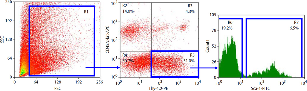
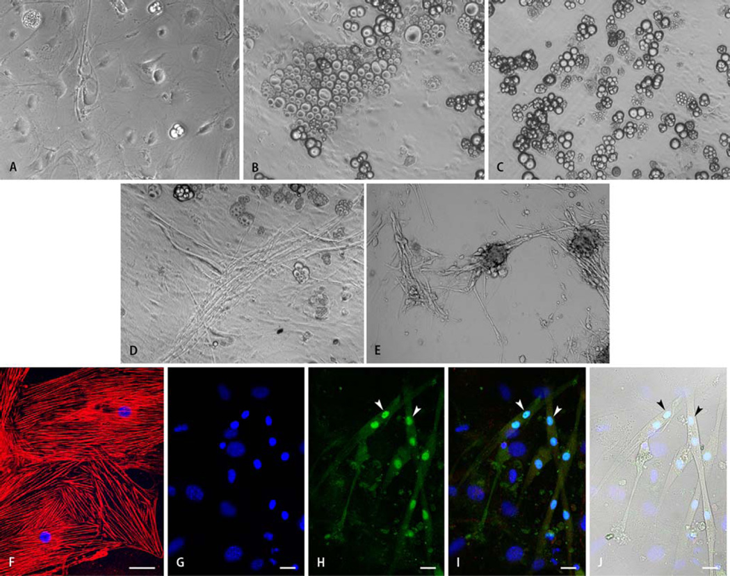
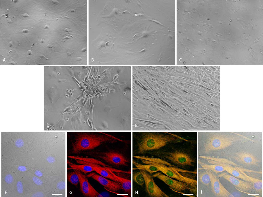
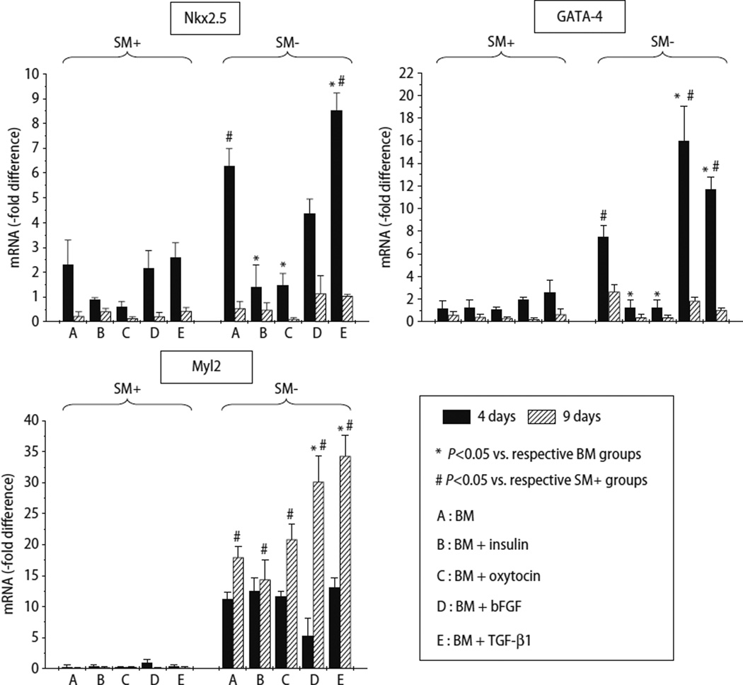
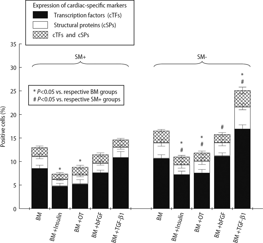
The optimal medium for cardiac differentiation of adult primitive cells remains to be established. We quantitatively compared the efficacy of IGF-1, dynorphin B, insulin, oxytocin, bFGF, and TGF-beta1 in inducing cardiomyogenic differentiation. Adult mouse skeletal muscle-derived Sca1+/CD45-/c-kit-/Thy-1+ (SM+) and Sca1-/CD45-/c-kit-/Thy-1+ (SM-) cells were cultured in basic medium (BM; DMEM, FBS, IGF-1, dynorphin B) alone and BM supplemented with insulin, oxytocin, bFGF, or TGF-beta1. Cardiac differentiation was evaluated by the expression of cardiac-specific markers at the mRNA (qRT-PCR) and protein (immunocytochemistry) levels. BM+TGF-beta1 upregulated mRNA expression of Nkx2.5 and GATA-4 after 4 days and Myl2 after 9 days. After 30 days, BM+TGF-beta1 induced the greatest extent of cardiac differentiation (by morphology and expression of cardiac markers) in SM- cells. We conclude that TGF-beta1 enhances cardiomyogenic differentiation in skeletal muscle-derived adult primitive cells. This strategy may be utilized to induce cardiac differentiation as well as to examine the cardiomyogenic potential of adult tissue-derived stem/progenitor cells.


Figures

Fig. 1
Fig. 1
Representative dot-plots show the expression (in percentage) of Sca-1 (FITC), CD45 (APC), c-kit (APC), and Thy-1.2 (PE) in skeletal muscle-derived cells. Blue lines indicate regions included into the sorting logic. From region 1 (R1), CD45-/c-kit- cells positive for Thy-1.2 are identified in region 5 (R5). Based on Sca-1 expression, cells from R5 are divided into Sca-1 +/CD45-/c-kit-/Thy-1 + (SM+) cells in region 7 (R7) and Sca-1-/CD45-/c-kit-/Thy-1+ (SM−) cells in region 6 (R6). The percentages in R6 and R7 relate to the total number of cells in R1. FSC forward scatter characteristics, SSC side scatter characteristics
Fig. 2
Fig. 2
Representative light microscopic images of SM+ cells after 30 days of differentiation demonstrating the effects of culture media composition on cellular morphology. Panels a–c show the heterogeneous (epithelial, endothelial, adipose, and others) morphology of SM+ cells cultured in basic medium (BM) containing IGF-1 and dynorphin B (a), and BM supplemented with insulin (b), or oxytocin (c). Exposure of SM+ cells to insulin or oxytocin increased differentiation into an adipose phenotype. Supplementation of BM with bFGF (d) or TGF-β1 (e) resulted in enhanced myotube formation from SM+ cells. Morphological observations were confirmed via immunostaining (f–j). Panel f shows differentiation of SM+ cells cultured in BM supplemented with insulin into epithelial cells positive for the epithelial cytoskeletal marker cytokeratin 17 (red). Panels g–j show representative images of SM+ cells stained for the cardiac-specific marker cardiac myosin heavy chain (i,j red), and skeletal muscle-specific transcription factor myogenin (h–j, green). Nuclei are stained with DAPI (f,g,i,j, blue). Myogenin expression was localized to nuclei (H,l, exemplified by arrowheads). Nuclei expressing myogenin (h–j, exemplified by arrowheads) belonged to cells negative for cardiac marker in red fluorescence (h,j) and exhibited morphology characteristic of myotubes (j). Scale bar = 40 µm (f) or 20 µm (g–j)
Fig. 3
Fig. 3
Representative light microscopic images of SM− cells after 30 days of differentiation demonstrating the effects of culture media composition on cellular morphology. Panels a–c show the predominant epithelial morphology of SM-cells cultured in basic medium (BM) containing IGF-1 and dynorphin B (a), and BM supplemented with insulin (b), or oxytocin (c). Scattered cells with adipose morphology were noted throughout the culture plates. Panels d and e show the increased frequency of spindle-shaped cells when SM− cells were exposed to BM supplemented with bFGF (d) or TGF-β1 (e). Cardiomyogenic differentiation was verified via immunostaining. Panels f–i show representative confocal microscopic images of SM− cells after 30 days of differentiation in BM supplemented with TGF-β1. Mononucleate spindle-shaped cells are identified in the transmission image (f). Cardiac differentiation is evidenced by positivity for cardiac-specific transcription factor (Nkx2.5h–i, green) and structural protein (cardiac myosin heavy chain, g–i, red). Panels h–i identify cells positive for both cardiac-specific transcription factor (in green) and structural protein (in red) resulting in yellow fluorescence. Nuclei are stained with DAPI (f,g,i, blue). Scale bar = 20 µm
Fig. 4
Fig. 4
Quantitative assessment of mRNA expression by qRT-PCR of cardiac-specific markers in SM+ and SM− cells after 4 and 9 days of culture in 5 different media. mRNA level is expressed as -fold change compared with freshly isolated unfractionated skeletal muscle-derived cells. Three independent experiments were performed. Data are mean ± SEM
Fig. 5
Fig. 5
Percentage of cells positive for cardiac-specific markers (Y axis), demonstrating the effects of different media (X axis) on cardiomyogenic differentiation of SM+ and SM− cells. Cells positive for cardiac transcription factors (cTFs) and/or structural proteins (cSPs) were counted and expressed as a percentage of total cells. The percentage of cells positive for cTFs is represented by solid bars, cSPs by white bars, and cells positive for both by cross-hatched bars. Cardiomyocytic differentiation was noted in both SM+ and SM− populations. However, compared with SM+ cells, SM− cells exhibited greater cardiomyogenic potential in all media. Supplementation of BM with TGF-β1 resulted in cardiomyocytic differentiation in the greatest fraction of cells in both groups

References

    1. Abdel-Latif A, Bolli R, Tleyjeh IM, Montori VM, Perin EC, Hornung CA, Zuba-Surma EK, Al-Mallah M, Dawn B. Adult bone marrow-derived cells for cardiac repair: a systematic review and meta-analysis. Arch Intern Med. 2007;167:989–997. - PubMed
    1. Antin PB, Yatskievych T, Dominguez JL, Chieffi P. Regulation of avian precardiac mesoderm development by insulin and insulin-like growth factors. J Cell Physiol. 1996;168:42–50. - PubMed
    1. Bani-Yaghoub M, Kendall SE, Moore DP, Bellum S, Cowling RA, Nikopoulos GN, Kubu CJ, Vary C, Verdi JM. Insulin acts as a myogenic differentiation signal for neural stem cells with multilineage differentiation potential. Development. 2004;131:4287–4298. - PubMed
    1. Barron M, Gao M, Lough J. Requirement for BMP and FGF signaling during cardiogenic induction in non-precardiac mesoderm is specific, transient, and cooperative. Dev Dyn. 2000;218:383–393. - PubMed
    1. Bassas L, Lesniak MA, Serrano J, Roth J, de Pablo F. Developmental regulation of insulin and type I insulin-like growth factor receptors and absence of type II receptors in chicken embryo tissues. Diabetes. 1988;37:637–644. - PubMed

Publication types

MeSH terms