. 2008 May 23:9:244.
doi: 10.1186/1471-2164-9-244.

Cloning, characterization, and expression of microRNAs from the Asian malaria mosquito, Anopheles stephensi

Affiliations

Cloning, characterization, and expression of microRNAs from the Asian malaria mosquito, Anopheles stephensi

Edward Andrew Mead et al. BMC Genomics. .

Abstract

Background: microRNAs (miRNAs) are non-coding RNAs that are now recognized as a major class of gene-regulating molecules widely distributed in metozoans and plants. miRNAs have been found to play important roles in apoptosis, cancer, development, differentiation, inflammation, longevity, and viral infection. There are a few reports describing miRNAs in the African malaria mosquito, Anopheles gambiae, on the basis of similarity to known miRNAs from other species. An. stephensi is the most important malaria vector in Asia and it is becoming a model Anopheline species for physiological and genetics studies.

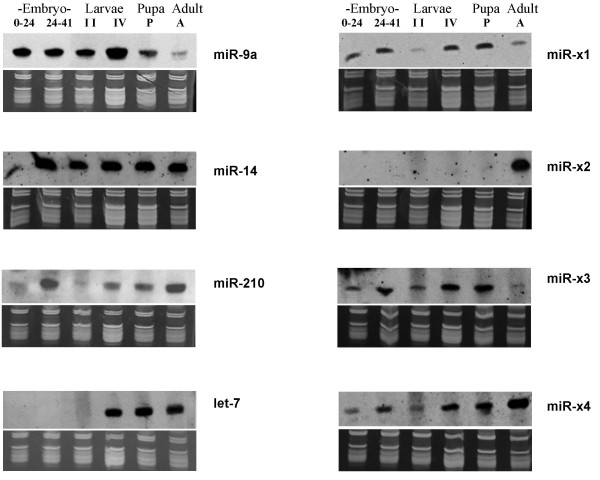
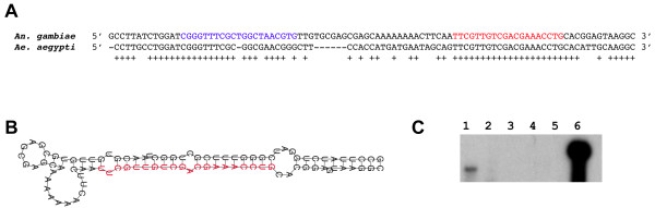
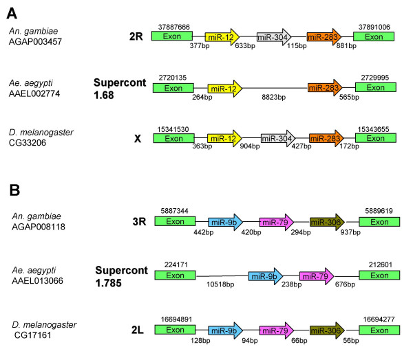
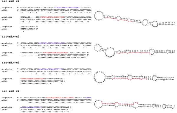
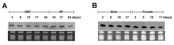
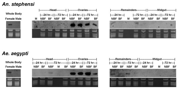
Results: We report the cloning and characterization of 27 distinct miRNAs from 17-day old An. stephensi female mosquitoes. Seventeen of the 27 miRNAs matched previously predicted An. gambiae miRNAs, offering the first experimental verification of miRNAs from mosquito species. Ten of the 27 are miRNAs previously unknown to mosquitoes, four of which did not match any known miRNAs in any organism. Twenty-five of the 27 Anopheles miRNAs had conserved sequences in the genome of a divergent relative, the yellow fever mosquito Aedes aegypti. Two clusters of miRNAs were found within introns of orthologous genes in An. gambiae, Ae. aegypti, and Drosophila melanogaster. Mature miRNAs were detected in An. stephensi for all of the nine selected miRNAs, including the four novel miRNAs (miR-x1- miR-x4), either by northern blot or by Ribonuclease Protection Assay. Expression profile analysis of eight of these miRNAs revealed distinct expression patterns from early embryo to adult stages in An. stephensi. In both An. stephensi and Ae. aegypti, the expression of miR-x2 was restricted to adult females and predominantly in the ovaries. A significant reduction of miR-x2 level was observed 72 hrs after a blood meal. Thus miR-x2 is likely involved in female reproduction and its function may be conserved among divergent mosquitoes. A mosquito homolog of miR-14, a regulator of longevity and apoptosis in D. melanogaster, represented 25% of all sequenced miRNA clones from 17-day old An. stephensi female mosquitoes. An. stephensi miR-14 displayed a relatively strong signal from late embryonic to adult stages. miR-14 expression is consistent during the adult lifespan regardless of age, sex, and blood feeding status. Thus miR-14 is likely important across all mosquito life stages.

Conclusion: This study provides experimental evidence for 23 conserved and four new microRNAs in An. stephensi mosquitoes. Comparisons between miRNA gene clusters in Anopheles and Aedes mosquitoes, and in D. melanogaster suggest the loss or significant change of two miRNA genes in Ae. aegypti. Expression profile analysis of eight miRNAs, including the four new miRNAs, revealed distinct patterns from early embryo to adult stages in An. stephensi. Further analysis showed that miR-x2 is likely involved in female reproduction and its function may be conserved among divergent mosquitoes. Consistent expression of miR-14 suggests that it is likely important across all mosquito life stages from embryos to aged adults. Understanding the functions of mosquito miRNAs will undoubtedly contribute to a better understanding of mosquito biology including longevity, reproduction, and mosquito-pathogen interactions, which are important to disease transmission.


Figures

Figure 1
Figure 1
Northern analysis of eight miRNAs across different developmental stages in An. stephensi. Shown here are eight northern blots performed using Dig-labeled miRCURY LNA probes designed for hybridization to either miR-14, let-7, miR-9a, miR-210, or to one of the four novel miRNAs (miR-x1–x4). The top panels are northern results and the bottom panels are RNA gels for verification of small ribosomal and tRNA integrity and equal loading of total RNA. ssDNA size markers (19 and 23 nts, not shown) were also visualized on the RNA gel for size estimation. Ten micrograms of total RNA for each sample were used. Developmental stages examined were early embryo (Embryo 0–24: 0–24 hrs after egg deposition), late embryo (Embryo 24–41: 24–41 hrs after egg deposition), intermediate and late larval stages (II and IV, respectively), Pupa (P), and Adult (A). To be consistent with our cloning experiment, 17-day old adult females were used in these northern experiments.
Figure 2
Figure 2
Analysis of ast-mir-76, a miRNA that was previously unknown in mosquitoes. The mature miRNA sequence was cloned from An. stephensi, and the hairpin precursor sequence was obtained from the An. gambiae genome assembly. A) Alignment of the pre-miRNA hairpins found in An. gambiae and Ae. aegypti. The mature miRNA is marked in red while miRNA* is marked in blue. Conserved nucleotides are indicated by a "+". B) Hairpin structure of An. gambiae mir-76. The mature miRNA predicted by miRscan (Table 2) is shown in red. C) RPA analysis of ast-mir-76. Lane 1, An. stephensi RNA with probe and digested; Lane 2, yeast RNA with probe and digested; Lane 3, yeast RNA without probe and digested; Lanes 4 and 5, empty lanes; Lane 6, undigested probe. A band of the correct size was only observed in An. stephensi total RNA samples (Lane 1). The size of the protected RNA product in lane 1 was estimated to be 24 nucleotides using markers as described in Figure 1. This size is as expected (the protected 20-nt long ast-miR-76 plus 4 undigested adenosines, see Methods).
Figure 3
Figure 3
Clustering of miRNAs genes. A) A miRNA gene cluster within an intron of a conserved gene of unknown function. The miRNA gene cluster contains miR-12, -283, and -304. B) A miRNA gene cluster within an intron of a gene coding for a serine-threonine kinase group protein. The miRNA gene cluster contains miR-9b, -79, and -306. Note that one miRNA was not found in the genome of Ae. aegypti in both panels. Species name and gene identification are provided at the left side of the figure. Chromosome or supercontig numbers are indicated right next to diagram depicting the miRNA gene clusters. Chromosomal or supercontig positions of the regions depicted are above the boxes showing the exons. miRNA genes are shown as open arrows. The distance between the miRNA genes and neighboring exons are indicated below the diagram. The figure is not drawn to scale. The exons shown in both panels are orthologous as indicated by conserved amino acid sequences.
Figure 4
Figure 4
Sequence alignment and predicted secondary structure of four novel miRNAs. Shown on the left are the sequence alignments between An. gambiae and Ae. aegypti miRNA precursor hairpins. Plus signs indicate conservation. The mature miRNA is marked in red while miRNA* is marked in blue. Note the perfect conservation of the mature miRNA (red), high conservation of the miRNA* sequence (blue), and lower conservation of the surrounding stem and loop structure, a hallmark conservation pattern of pre-miRNAs. Shown on the right are the predicted secondary structures of corresponding An. gambiae miRNA hairpins. The mature miRNA is marked in red on the hairpin.
Figure 5
Figure 5
miR-14 expression across An. stephensi adult lifespan. Shown here are northern blots performed using Dig-labeled miRCURY LNA probes designed for hybridization to miR-14. The top panel is the northern result and the bottom panel is a corresponding RNA gel for verification of small ribosomal and tRNA integrity and equal loading of total RNA. ssDNA size markers (19 and 23 nts, not shown) were also visualized on the RNA gel for size estimation. Ten micrograms of total RNA for each sample were used. A) miR-14 expression in An. stephensi adult females fed with either sugar water (NBF, non-bloodfed) or blood meal (BF, bloodfed). The samples were 3, 5, 10, 17, and 24 day old adult females that were maintained on sugar water as well as adult females that were fed on blood on day 5 after emergence and collected at day 10, 17, and 24. Bloodfed females were allowed to oviposit two days after the blood meal. B) miR-14 expression in An. stephensi males and NBF females between 3–17 days of age. We did not extend the comparative analysis to 24 days post emergence because the majority of males do not survive that long.
Figure 6
Figure 6
Expression of miR-x2 in An. stephensi and Ae. aegypti: sex-specificity, tissue distribution and the impact of blood feeding. Shown here are northern blots performed using Dig-labeled miRCURY LNA probes designed for hybridization to miR-x2. The top panels are northern results and the bottom panels are RNA gels for verification of small ribosomal and tRNA integrity and equal loading of total RNA. ssDNA size markers (19 and 23 nts, not shown) were also visualized on the RNA gel for size estimation. On the left panel for each species, a comparison between 5-day old adult male and 5-day old non-bloodfed female is shown. Ten micrograms of total RNA isolated from the whole mosquitoes were used. The middle and right panels are comparisons between adult female tissues or body parts in each species. Tissues used were Heads, Ovaries, Midguts, and Remainders. There were four samples for each tissue: BF, tissue sample from bloodfed females at 24 and 72 hrs post-bloodfeeding; NBF, tissue sample from non-bloodfed (sugar-fed) females at equivalent time points compared to the blood-fed samples. Five micrograms of total RNA for each sample were used. The markers lane is designated with an 'M' although the markers are not within the gel image panel because they are below the size of the ribosomal and tRNA.

References

    1. Miska EA. How microRNAs control cell division, differentiation and death. Curr Opin Genet Dev. 2005;15:563–568. doi: 10.1016/j.gde.2005.08.005. - DOI - PubMed
    1. Du T, Zamore PD. Beginning to understand microRNA function. Cell Research. 2007;17:661–663. doi: 10.1038/cr.2007.67. - DOI - PubMed
    1. Bartel DP. MicroRNAs: genomics, biogenesis, mechanism, and function. Cell. 2004;116:281–297. doi: 10.1016/S0092-8674(04)00045-5. - DOI - PubMed
    1. Pasquinelli AE, Hunter S, Bracht J. MicroRNAs: a developing story. Curr Opin Genet Dev. 2005;15:200–205. doi: 10.1016/j.gde.2005.01.002. - DOI - PubMed
    1. Zhou X, Ruan J, Wang G, Zhang W. Characterization and identification of MicroRNA core promoters in four model species. PLoS Comput Biol. 2007;3:e37. doi: 10.1371/journal.pcbi.0030037. - DOI - PMC - PubMed

Publication types

MeSH terms

LinkOut - more resources