. 2008 Aug 15;45(4):443-52.
doi: 10.1016/j.freeradbiomed.2008.04.038. Epub 2008 May 3.

Oxidative stress and modification of synaptic proteins in hippocampus after traumatic brain injury

Affiliations

Oxidative stress and modification of synaptic proteins in hippocampus after traumatic brain injury

Mubeen A Ansari et al. Free Radic Biol Med. .

Abstract

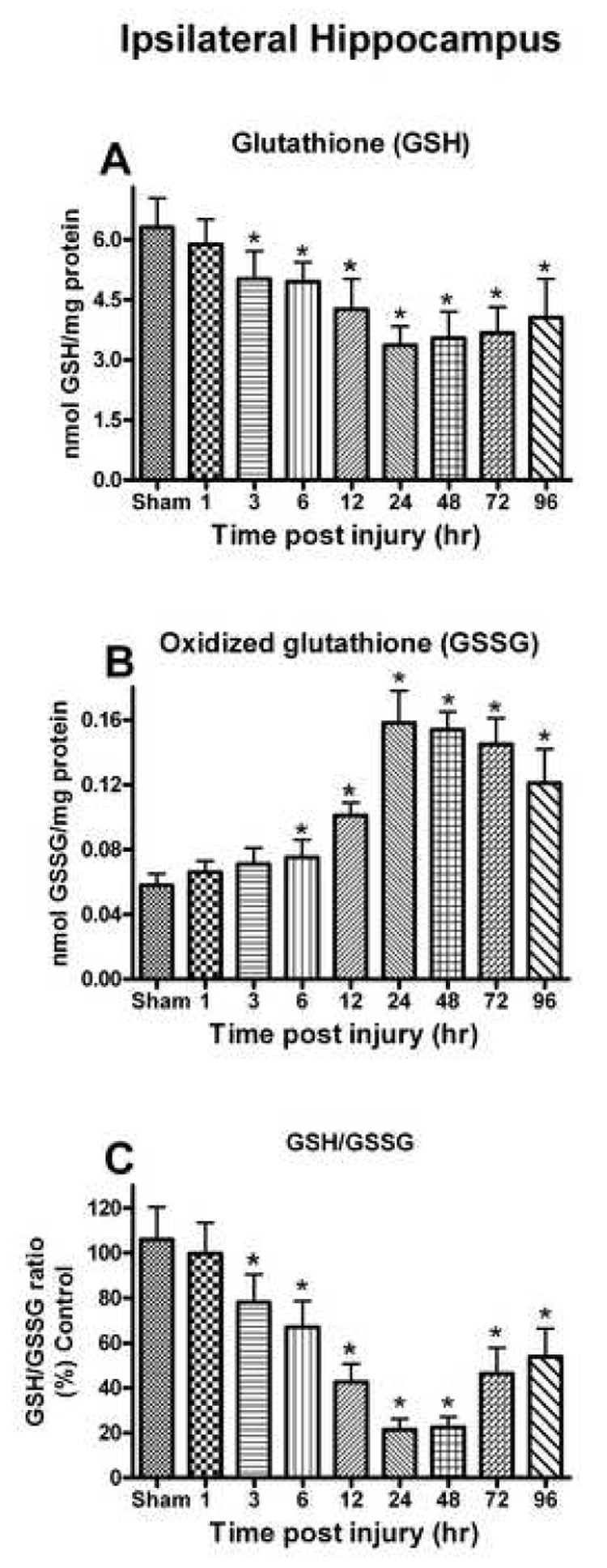
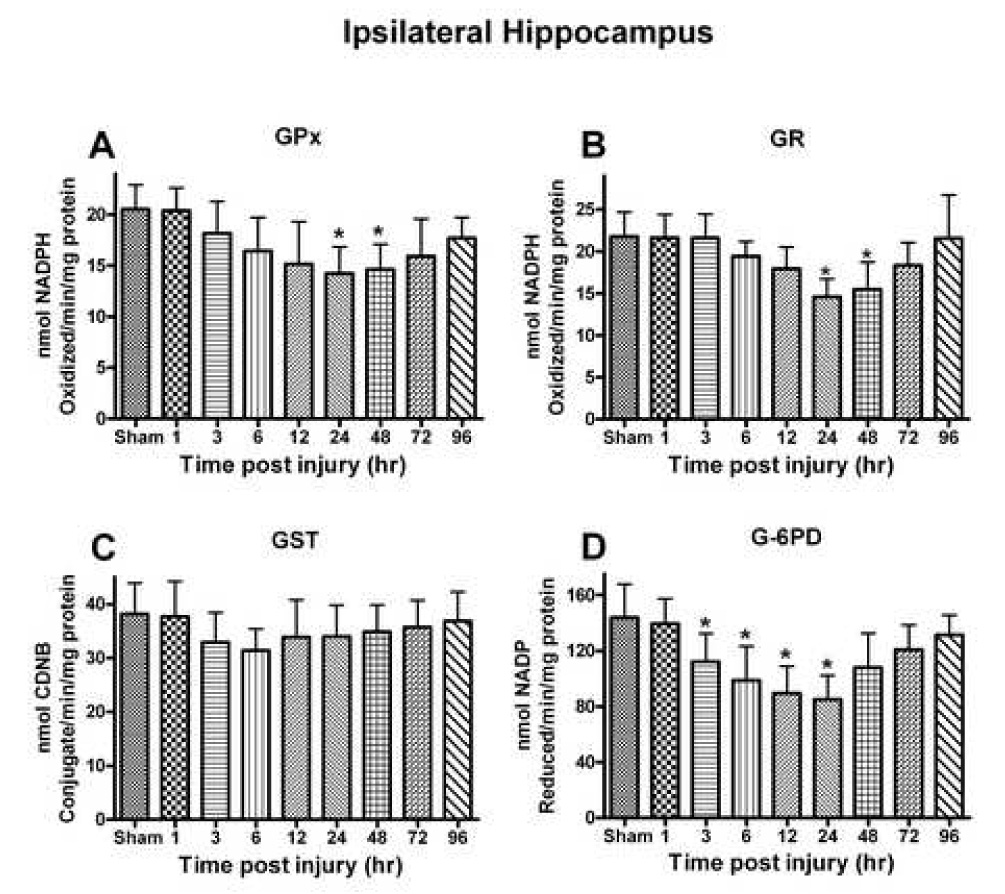
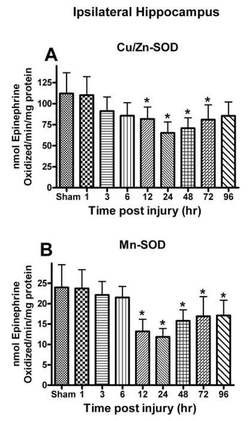
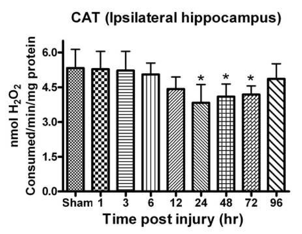
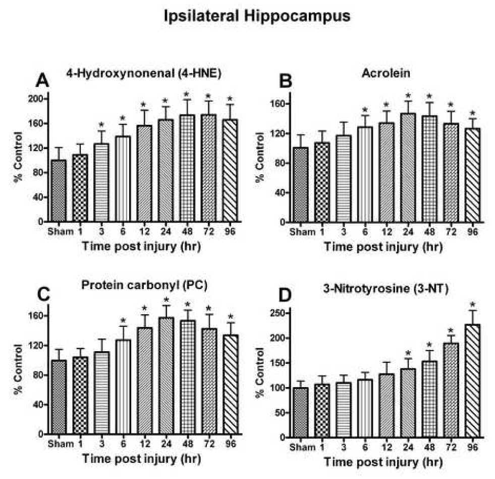
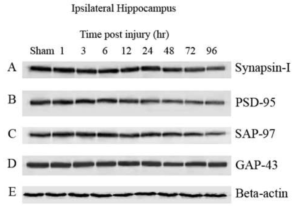
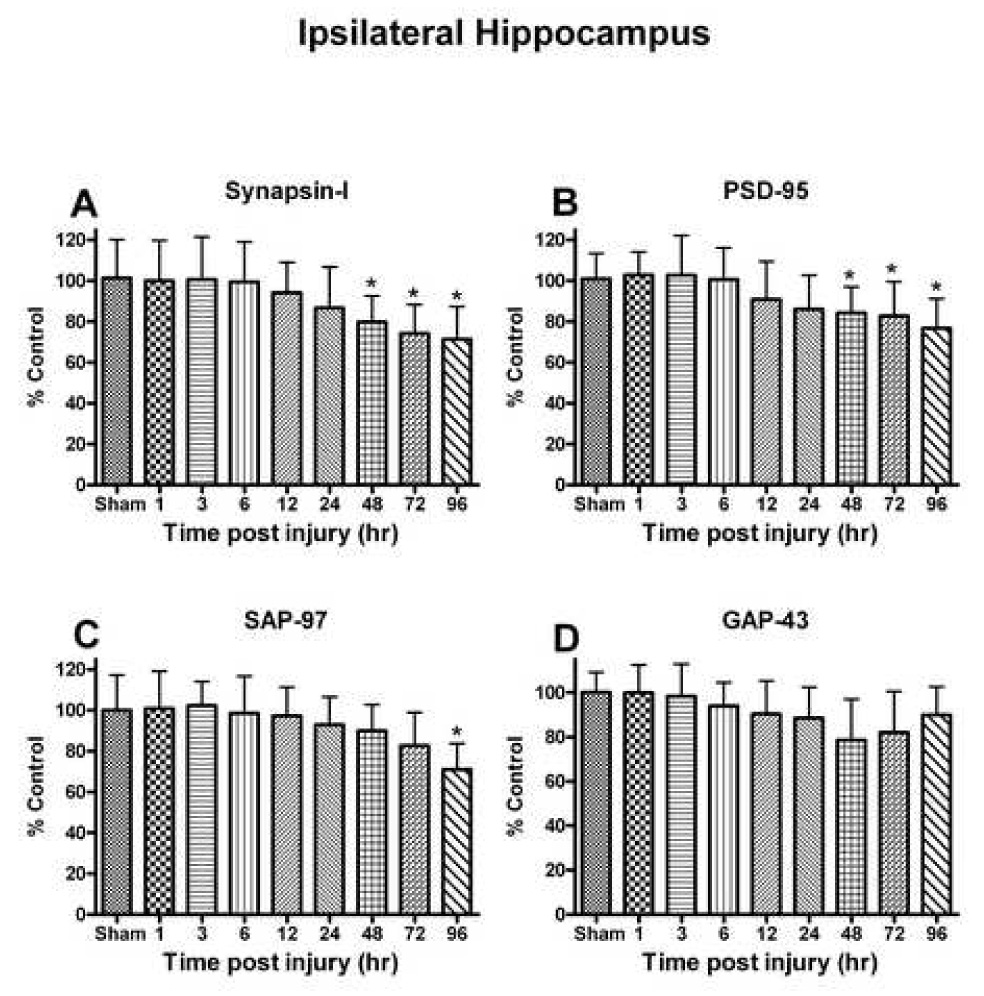
Oxidative stress, an imbalance between oxidants and antioxidants, contributes to the pathogenesis of traumatic brain injury (TBI). Oxidative neurodegeneration is a key mediator of exacerbated morphological responses and deficits in behavioral recoveries. The present study assessed early hippocampal sequential imbalance to possibly enhance antioxidant therapy. Young adult male Sprague-Dawley rats were subjected to a unilateral moderate cortical contusion. At various times post-TBI, animals were killed and the hippocampus was analyzed for antioxidants (GSH, GSSG, glutathione peroxidase, glutathione reductase, glutathione-S-transferase, glucose-6-phosphate dehydrogenase, superoxide dismutase, and catalase) and oxidants (acrolein, 4-hydroxynonenal, protein carbonyl, and 3-nitrotyrosine). Synaptic markers (synapsin I, postsynaptic density protein 95, synapse-associated protein 97, growth-associated protein 43) were also analyzed. All values were compared with those for sham-operated animals. Significant time-dependent changes in antioxidants were observed as early as 3 h posttrauma and paralleled increases in oxidants (4-hydroxynonenal, acrolein, and protein carbonyl), with peak values obtained at 24-48 h. Time-dependent changes in synaptic proteins (synapsin I, postsynaptic density protein 95, and synapse-associated protein 97) occurred well after levels of oxidants peaked. These results indicate that depletion of antioxidant systems following trauma could adversely affect synaptic function and plasticity. Early onset of oxidative stress suggests that the initial therapeutic window following TBI appears to be relatively short, and it may be necessary to stagger selective types of antioxidant therapy to target specific oxidative components.


Figures

Fig. 1
Fig. 1
Changes in the GSH system (GSH, GSSG and GSH/GSSG) after a unilateral cortical contusion in male SD rats. (A) As early as 3h post trauma, levels of GSH were significantly decreased and depletion of GSH levels persisted for up to 96h. (B) The levels of GSSG were significantly increased at 6h and remained significantly elevated for up to 96h. (C) At the 3h post injury, levels of GSH/GSSG were significantly altered and this depletion in ratio remained significant at 96h. Each bar represents the group mean ± SD of six animals/group. *p < 0.05 versus sham.
Fig. 2
Fig. 2
Several different antioxidant enzymes were quantitatively assessed following a moderate unilateral cortical contusion. (A) Glutathione peroxidase (GPx) activity demonstrated a time-dependent change with significant reductions observed at 24–48h post injury. (B) Glutathione reductase (GR) activity demonstrated a time-dependent decline that mirrored that observed for GPx. (C) Glutathione-S-transferase (GST) activity had non-significant depletion very early following the trauma and remained relatively unchanged. (D) Glucose-6-phosphate dehydrogenase (G-6PD) activity altered significantly at 3h with maximum depletion by 24h, eventually returning to control levels at 96h post injury. Each bar represents the group mean ± SD of six animals/group. *p < 0.05 versus sham.
Fig. 3
Fig. 3
(A) Cu/Zn-SOD and (B) Mn-SOD activity in the ipsilateral hippocampus was affected post TBI. Cu/Zn-SOD activity declined at 12h following the trauma and approached sham levels by 96h post injury. Mn-SOD activity demonstrated a time-dependent decline that was greater than that observed for Cu/Zn-SOD and failed to return to control levels. Each bar represents the group mean ± SD of six animals/group. *p < 0.05 versus sham.
Fig. 4
Fig. 4
CAT activity in the hippocampus significantly declined following a unilateral cortical contusion. Significant depletion was observed by 24h post TBI with a return to control levels by 96h. Enzyme activity was measured with H2O2. Each bar represents group mean ± SD of six animals/group. *p < 0.05 versus sham.
Fig. 5
Fig. 5
The effect of a moderate unilateral cortical contusion on lipid peroxidation and protein modification at different time points post TBI. (A) 4-HNE levels demonstrated a time-dependent increase in the ipsilateral hippocampus following trauma and remained elevated at 96h post trauma. (B) Acrolein levels were increased significantly as early as 6h following trauma and never returned to control levels by 96h. (C) PC levels demonstrated a time-dependent change that mirrored that observed for acrolein with significant increase observed as early as 6h post trauma. (D) Changes in 3-NT levels were delayed in response to the trauma with a significant increase first observed at 24h that failed to returne to control levels by 96h post injury. Each bar represents the group mean ± SD of six animals/group. *p < 0.05 versus sham.
Fig. 6
Fig. 6
Synaptic proteins in the ipsilateral hippocampus were significantly affected following a unilateral cortical contusion. Hippocampal sample from six animals/group were processed for immunobloting followed by Western-blot. Immunoblotes were developed with 5- Bromo-4-chloro-3-indolyl phosphate/Nitro blue tetrazolium (alkaline phosphatase substrate; SIGMA FAST™ BCIP/NBT) and densities of bands were analyzed using Scion Image. Synapsin-I, PSD-95, and synapse associated protein -97 (SAP-97) demonstrated a time-dependent decrease in signal. Growth associated protein-43 (GAP-43) and Beta-actin failed to demonstrate any time-dependent decline in levels in the ipsilateral hippocampus.
Fig. 7
Fig. 7
(A) Pre-synaptic marker protein synapsin-I levels in the ipsilateral hippocampus declined following a unilateral cortical contusion and failed to return to sham injury levels by 96h. (B) Post-synaptic density protein -95 (PSD-95) levels mirrored the changes observed with synapsin-I and remained significantly decreased by 96h. (C) SAP-97 levels demonstrated a significant decline following the trauma at 96h. (D) Growth associated protein-43 (GAP-43) failed to demonstrate a significant change following TBI up to 96hr. Each bar represents the group mean ± SD of six animals/group. *p < 0.05 versus sham.
Fig. 8
Fig. 8
The total number of Fluoro-Jade B positive neurons in the different regions of the ipsilateral hippocampus following a unilateral cortical contusion. As early as 1h following TBI, significant number of FJB-positive neurons were observed in the dentate gyrus granule cell layer and CA3. The CA1 pyramidal cells showed the least number of positive cells with significant increase observed at 6h post contusion. Peak values were observed at 24h with significant number of FJB-positive cells still present at 7 day post trauma. Each bar represents the group mean ± SD. *p < 0.05 versus contralateral hippocampus as sham. 24h, 48h and 7 day data included from earlier published our lab findings (Anderson et al., 2005).
Fig. 9
Fig. 9
Ipsilateral coronal section through the hippocampal dentate gyrus and CA3 region at 24h following a unilateral cortical cortical contusion. Sections were stained with flour-Jade B (FJB) an anionic fluorescein derivative that specifically identifies degenerating neurons. Stereological techniques were used to determine the total number of FJB-positive neurons in different subregions of the hippocampal formation. Calibration bar = 500um

References

    1. Biegon A, Fry PA, Paden CM, Alexandrovich A, Tsenter J, Shohami E. Dynamic changes in N-methyl-D-aspartate receptors after closed head injury in mice: Implications for treatment of neurological and cognitive deficits. Proc. Natl. Acad. Sci. U. S. A. 2004;101:5117–5122. - PMC - PubMed
    1. Sullivan PG, Keller JN, Mattson MP, Scheff SW. Traumatic brain injury alters synaptic homeostasis: implications for impaired mitochondrial and transport function. J. Neurotrauma. 1998;15:789–798. - PubMed
    1. Engle MR, Singh SP, Czernik PJ, Gaddy D, Montague DC, Ceci JD, Yang Y, Awasthi S, Awasthi YC, Zimniak P. Physiological role of mGSTA4-4, a glutathione S-transferase metabolizing 4-hydroxynonenal: generation and analysis of mGsta4 null mouse. Toxicol. Appl. Pharmacol. 2004;194:296–308. - PubMed
    1. Shao C, Roberts KN, Markesbery WR, Scheff SW, Lovell MA. Oxidative stress in head trauma in aging. Free Radic. Biol. Med. 2006;41:77–85. - PubMed
    1. Ozdemir D, Uysal N, Gonenc S, Acikgoz O, Sonmez A, Topcu A, Ozdemir N, Duman M, Semin I, Ozkan H. Effect of melatonin on brain oxidative damage induced by traumatic brain injury in immature rats. Physiol. Res. 2005;54:631–637. - PubMed

Publication types