

Ophthalmic manifestations, cytology, immunohistochemistry, and molecular analysis of intraocular metastatic T-cell lymphoma: report of a case and review of the literature
- PMID: 18501272
- PMCID: PMC2474568
- DOI: 10.1016/j.survophthal.2008.02.004
Ophthalmic manifestations, cytology, immunohistochemistry, and molecular analysis of intraocular metastatic T-cell lymphoma: report of a case and review of the literature
Abstract
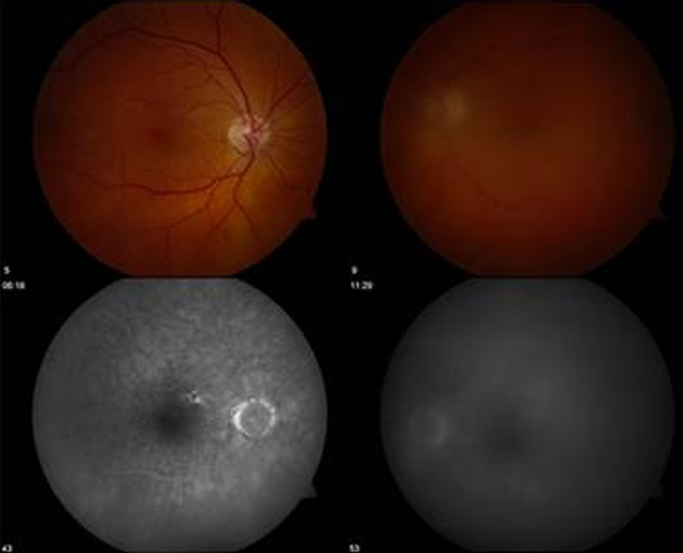
We report a case of T-cell lymphoma metastatic to the eye, with an accompanying review of the literature. A 78-year-old white male with bilateral vitritis was diagnosed with primary cutaneous peripheral T-cell lymphoma unspecified, via vitreous biopsy. The tumor was found to be clonally related to the prior cutaneous malignancy using cytology, immunophenotyping, and molecular analysis. The vast majority of primary intraocular lymphomas are malignant B-cells, whereas intraocular T-cell lymphomas are uncommon. This case demonstrates the utility of immunophenotyping and molecular analysis with microdissection and polymerase chain reaction, as critical adjunctive studies, in patients presenting with a masquerade syndrome, and later diagnosed with T-cell intraocular lymphomas. Vitreo-retinal without uveal involvement in this case, similar to many ocular metastatic T-cell lymphomas reported in the literature, is particularly intriguing because the uvea, not retina, is the typical ocular tissue involvement in the majority of metastatic B-cell lymphomas.
Figures

References
-
- Berenbom A, Davila RM, Lin HS, et al. Treatment outcomes for primary intraocular lymphoma: implications for external beam radiotherapy. Eye. 2007;21:1198–1201. - PubMed
-
- Bhatia S, Paulino AC, Buatti JM, et al. Curative radiotherapy for primary orbital lymphoma. Int J Radiat Oncol Biol Phys. 2002;54:818–823. - PubMed
-
- Buggage RR. Ocular manifestations of human T-cell lymphotropic virus type 1 infection. Curr Opin Ophthalmol. 2003;14:420–425. - PubMed
-
- Carlton WC, Hutchinson AK, Grossniklaus HE. Ocular tumors in animals and humans. Ames: Iowa State Press; 2002. Ocular lymphoid proliferations; pp. 379–413.
-
- Chan CC, Shen D, Nussenblatt RB, et al. Detection of molecular changes in primary intraocular lymphoma by microdissection and polymerase chain reaction. Diagn Mol Pathol. 1998;7:63–64. - PubMed
Publication types
MeSH terms
Substances
Grants and funding
LinkOut - more resources
Full Text Sources
Medical
