. 2008 Jun;86(6):867-78.
doi: 10.1016/j.exer.2008.01.002. Epub 2008 Jan 12.

Glucocorticoids induce transactivation of tight junction genes occludin and claudin-5 in retinal endothelial cells via a novel cis-element

Affiliations

Glucocorticoids induce transactivation of tight junction genes occludin and claudin-5 in retinal endothelial cells via a novel cis-element

Edward A Felinski et al. Exp Eye Res. 2008 Jun.

Abstract

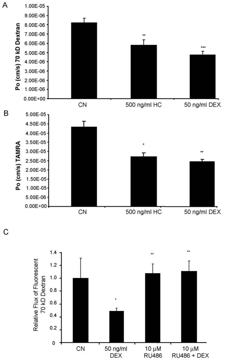
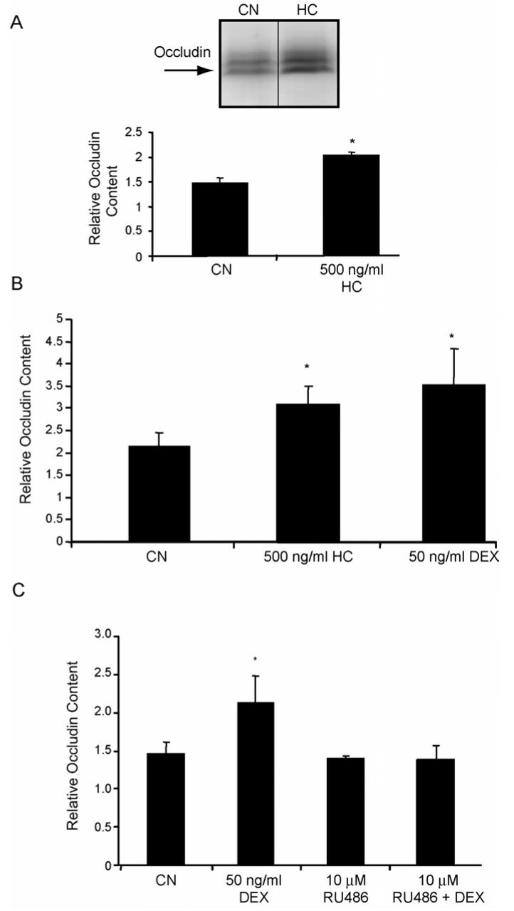
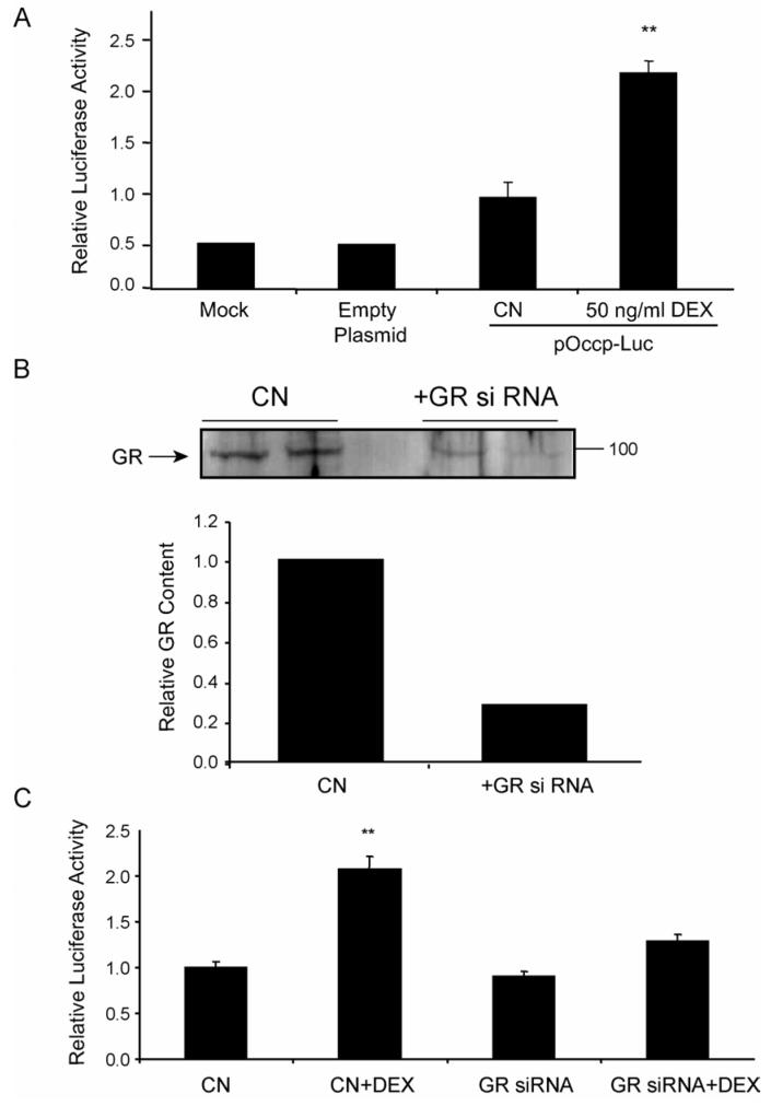
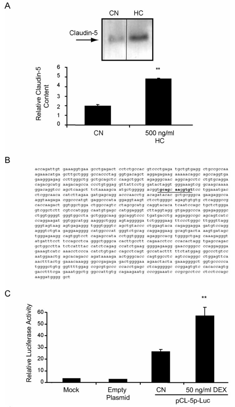
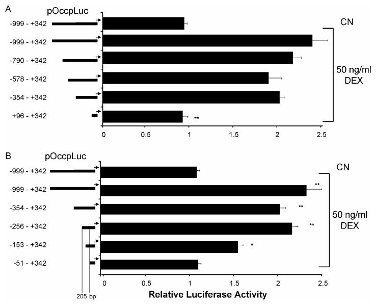
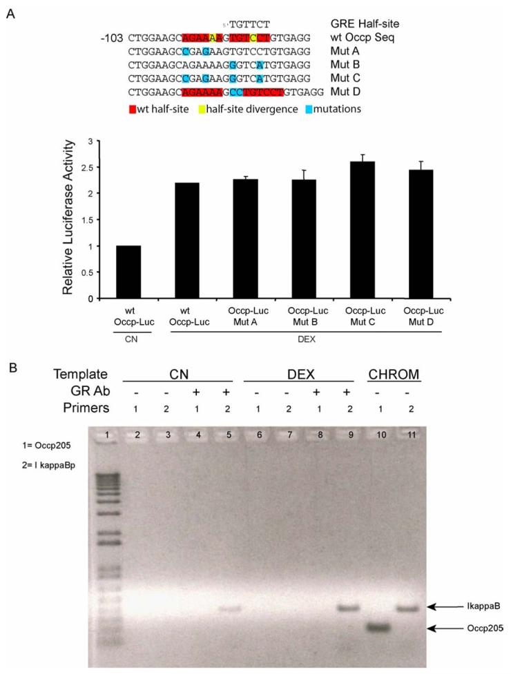
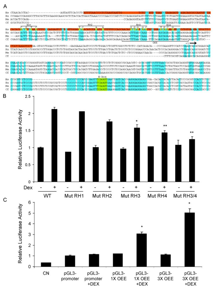
Tight junctions between vascular endothelial cells help to create the blood-brain and blood-retinal barriers. Breakdown of the retinal tight junction complex is problematic in several disease states including diabetic retinopathy. Glucocorticoids can restore and/or preserve the endothelial barrier to paracellular permeability, although the mechanism remains unclear. We show that glucocorticoid treatment of primary retinal endothelial cells increases content of the tight junction proteins occludin and claudin-5, co-incident with an increase in barrier properties of endothelial monolayers. The glucocorticoid receptor antagonist RU486 reverses both the glucocorticoid-stimulated increase in occludin content and the increase in barrier properties. Transcriptional activity from the human occludin and claudin-5 promoters increases in retinal endothelial cells upon glucocorticoid treatment, and is dependent on the glucocorticoid receptor (GR) as demonstrated by siRNA. Deletion analysis of the occludin promoter reveals a 205bp sequence responsible for the glucocorticoid response. However, this region does not possess a canonical glucocorticoid response element and does not bind to the GR in a chromatin immunoprecipitation (ChIP) assay. Mutational analysis of this region revealed a novel 40bp occludin enhancer element (OEE), containing two highly conserved regions of 10 and 13 base pairs, that is both necessary and sufficient for glucocorticoid-induced gene expression in retinal endothelial cells. These data suggest a novel mechanism for glucocorticoid induction of vascular endothelial barrier properties through increased occludin and claudin-5 gene expression.


Figures

Fig. 1
Fig. 1
Glucocorticoids regulate endothelial barrier properties. HREC (A&B) or BREC (C) were grown to confluence on 0.4 μm Transwell filters then treated with GC as indicated. (C) Cells were pretreated with 10 μM RU486 for 1 hour prior to GC treatment. After 24 hours, permeability of 70 kDa RITC-dextran (A&C) or 467 Dalton TAMRA (B) was determined as described. All graphs represent the mean +/- s.e.m. with analysis by ANOVA with Tukey post-test (n=4), * p < 0.05, ** p < 0.01, *** p < 0.001.
Fig. 2
Fig. 2
Glucocorticoids increase the content of occludin protein in retinal endothelial cells through a transactivation mechanism. BREC (A&C) or HREC (B) were grown to confluence then switched to media without serum (CN) or without serum plus 500 ng/ml hydrocortisone (HC) or 50 ng/ml dexamethasone (DEX) for 24 hours. (C) BREC were pretreated for 1 hour with 10 μM RU486 prior to Dex treatment. Whole-cell lysates were made in urea extraction buffer and western blots were probed with a primary antibody against occludin and quantified. Representative blots are shown with n=3. Graphs represent the mean +/- s.e.m. with analysis by ANOVA with Tukey post-test. * p < 0.05.
Fig. 3
Fig. 3
Occludin promoter activity is increased with glucocorticoid treatment dependent on the GR. (A) BREC were co-transfected with pGL3-Luc (empty Plasmid) or pOccp-Luc together with pRL-SV, treated with 50 ng/ml Dex for 24 hours and relative luciferase activity was determined. (B) HREC were transfected with siRNA specific for the human GR, cells were grown for 48 hours then cell lysates were assayed for GR by western blot and quantified. (C) HREC were cotransfected with GR siRNA and pOccp-Luc and treated with 50 ng/ml Dex for 24 hrs as indicated. Relative luciferase activity was determined as above. Data represents two independent experiments with n=6. Graphs represent the mean +/- s.e.m. with analysis by ANOVA with Tukey post-test ** p < 0.01.
Fig. 4
Fig. 4
Glucocorticoids increase the content of claudin-5 protein and the activity of the claudin-5 promoter in retinal endothelial cells. BREC were grown to confluence then switched to media without serum (CN) or without serum plus 500 ng/ml hydrocortisone (HC) for 24 hours. Whole-cell lysates were made in urea extraction buffer and western blots were probed with a primary antibody against claudin-5 and quantified. (B) Sequence of the 1500 bp upstream claudin-5 promoter sequence (sequence with homology to occludin RH4 highlighted). (C) BREC were transfected with pCL-5p-Luc and assayed for luciferase activity as above. Data represents three independent experiments with n=9. Graphs represent the mean +/- s.e.m. with analysis by ANOVA with Tukey post-test **p < 0.01.
Fig. 5
Fig. 5
The occludin promoter contains a 205 bp minimal glucocorticoid-responsive sequence. The indicated truncation mutants of the occludin promoter were synthesized using site-directed mutagenesis and used to transfect BREC. After 24 hours, cells were treated with 50 ng/ml Dex for 24 hrs. Relative luciferase activiy was measured and corrected for Renilla luciferase. Graphs represent the mean +/- s.e.m. with analysis by ANOVA with Tukey post-test of two independent experiments with n=6, ** p < 0.01.
Fig. 6
Fig. 6
GR does not directly bind the glucocorticoid responsive region of the occludin promoter. (A) Potential GRE half-sites are not necessary for the glucocorticoid response. Potential GRE half-sites within the occludin promoter 205 bp GC-responsive sequence identified by a cis-element search were mutated as indicated and used to transfect BREC cells as above. Transfected cells were treated with 50 ng/ml Dex and a dual luciferase assay was performed as described, n=3. (B) Confluent HREC were treated with 50 ng/ml Dex. A chromatin immunoprecipitation (ChIP) experiment was performed using primary antibody against the GR. Primers specific for the 205 bp GC-responsive fragment of the occludin promoter (primer set 1) or for a 400 bp GC-responsive fragment of the Iκ-B promoter (primer set 2) were used to perform PCR reactions from the precipitated DNA. CHROM is unprecipitated chromatin samples as a control for PCR. Graph represents the mean +/- s.e.m. with analysis by ANOVA with Tukey post-test of two independent experiments with n=6.
Fig. 7
Fig. 7
Identification of a novel occludin enhancer element (OEE) that responds to glucocorticoids in the occludin promoter. (A) Alignment of the occludin promoter from four mammalian species showing the 205 bp GC-responsive region in human (red) and bases conserved across all four species (blue). Point mutations made in the four regions of homology (RH1-4) are shown in yellow. (B) The indicated point mutants of Occp-Luc were used to transfect BREC and cells were treated with 50 ng/ml Dex for 24 hours. Relative luciferase activiy was measured and corrected for Renilla luciferase. (C) The occludin enhancer element (OEE) sequence indicated in (A) was subcloned as a monomer (pGL3-1XOEE) or in triplicate (pGL3-3XOEE) into the SV40 core promoter plasmid pGL3-promoter and used to transfect BREC as above. Graphs represent the mean +/- s.e.m. of two independent experiments with n=6 with analysis by ANOVA with Tukey post-test, * p < 0.05, ** p < 0.01.

References

    1. Aiello LP, Gardner TW, King GL, Blankenship G, Cavallerano JD, Ferris FL, 3rd, Klein R. Diabetic retinopathy. Diabetes Care. 1998;21:143–56. - PubMed
    1. Aijaz S, Balda MS, Matter K. Tight junctions: molecular architecture and function. Int Rev Cytol. 2006;248:261–98. - PubMed
    1. Anderson JM, Stevenson BR, Jesaitis LA, Goodenough DA, Mooseker MS. Characterization of ZO-1, a protein component of the tight junction from mouse liver and Madin-Darby canine kidney cells. J Cell Biol. 1988;106:1141–9. - PMC - PubMed
    1. Antonetti DA, Barber AJ, Khin S, Lieth E, Tarbell JM, Gardner TW. Vascular permeability in experimental diabetes is associated with reduced endothelial occludin content: vascular endothelial growth factor decreases occludin in retinal endothelial cells. Penn State Retina Research Group. Diabetes. 1998;47:1953–9. - PubMed
    1. Antonetti DA, Wolpert EB. Isolation and characterization of retinal endothelial cells. Methods Mol Med. 2003;89:365–74. - PubMed

Publication types

MeSH terms