Review
. 2008 Jul-Aug;1777(7-8):1001-19.
doi: 10.1016/j.bbabio.2008.04.037. Epub 2008 May 1.

The Q-cycle reviewed: How well does a monomeric mechanism of the bc(1) complex account for the function of a dimeric complex?

Affiliations
Review

The Q-cycle reviewed: How well does a monomeric mechanism of the bc(1) complex account for the function of a dimeric complex?

Antony R Crofts et al. Biochim Biophys Acta. 2008 Jul-Aug.

Abstract

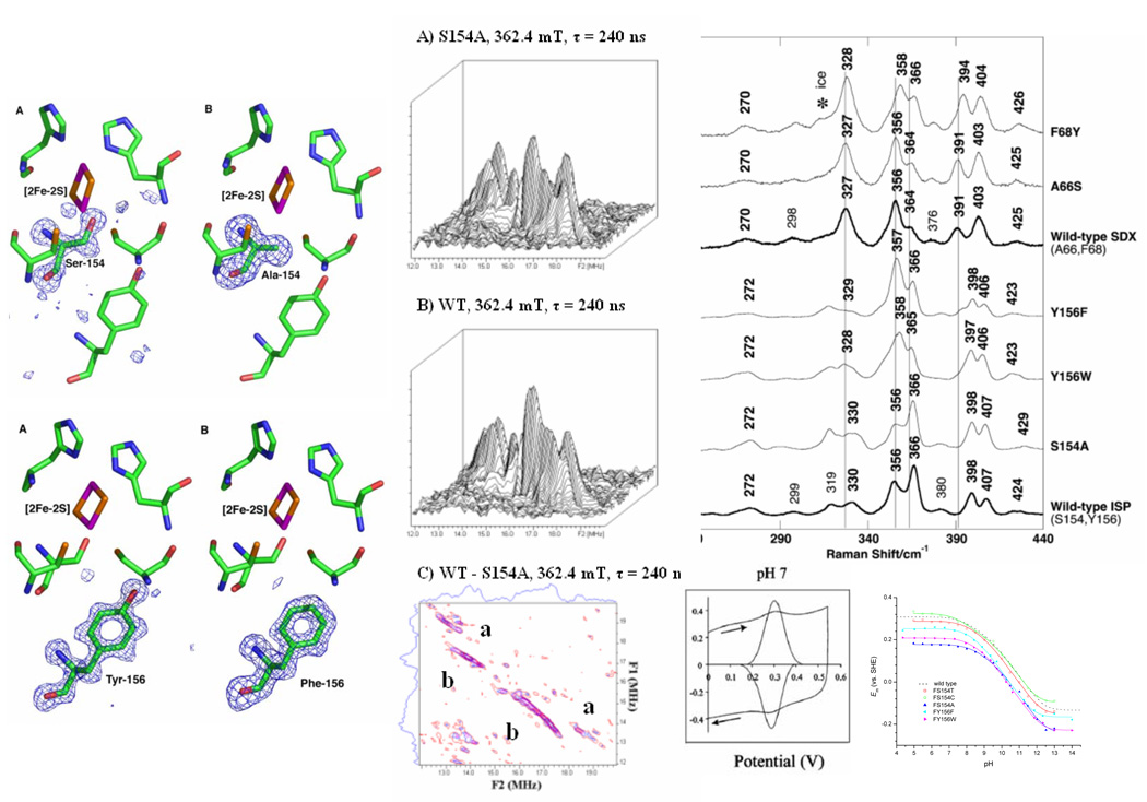
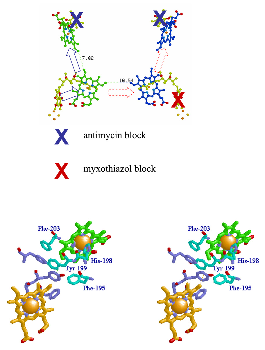
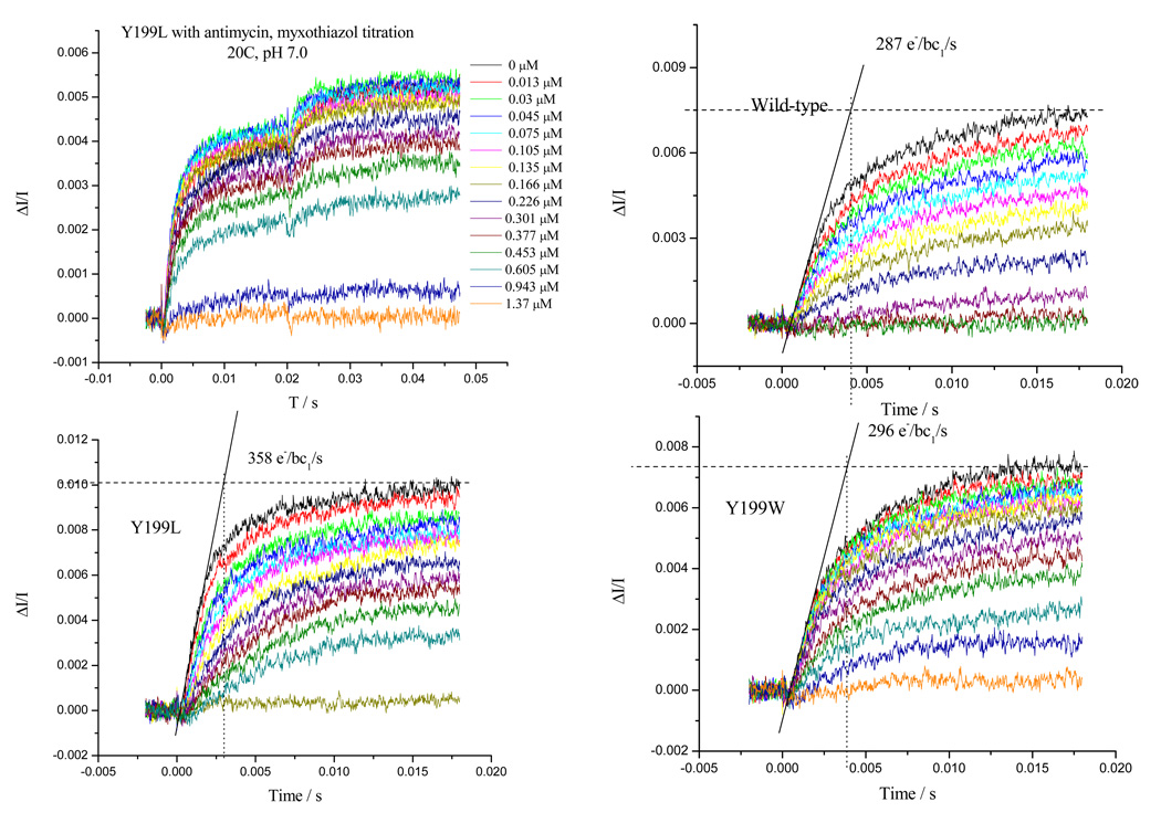
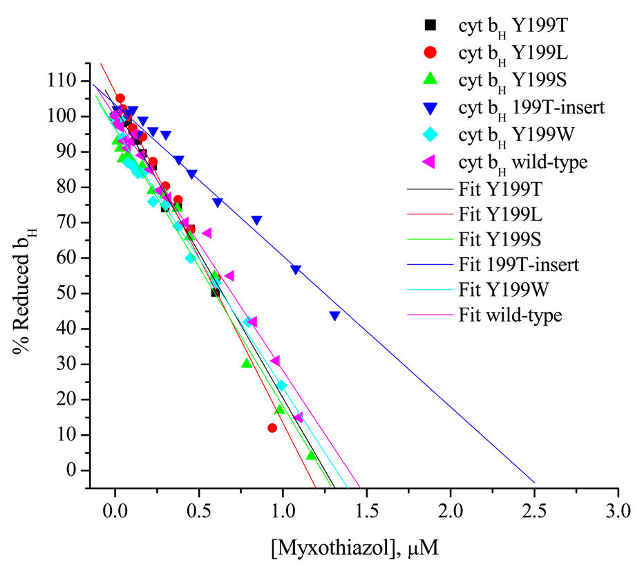
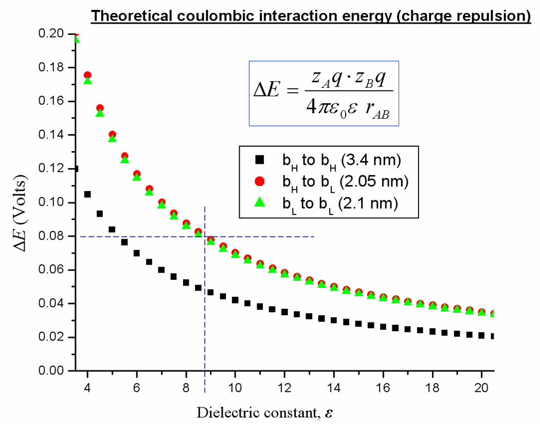
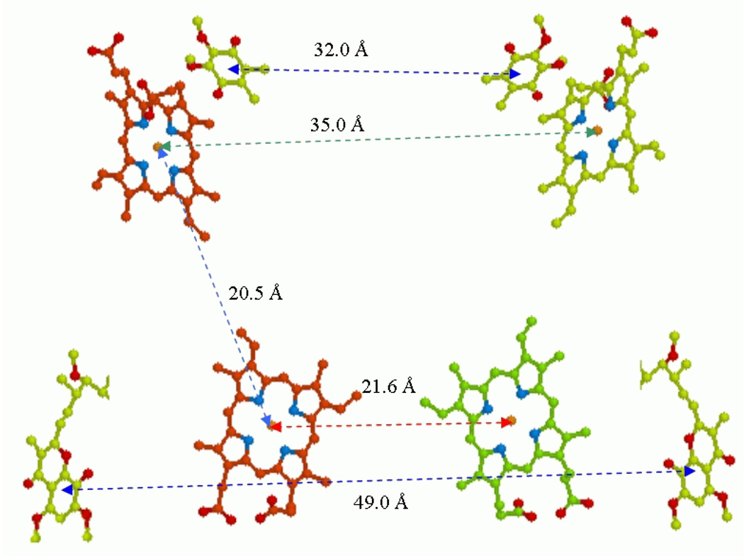
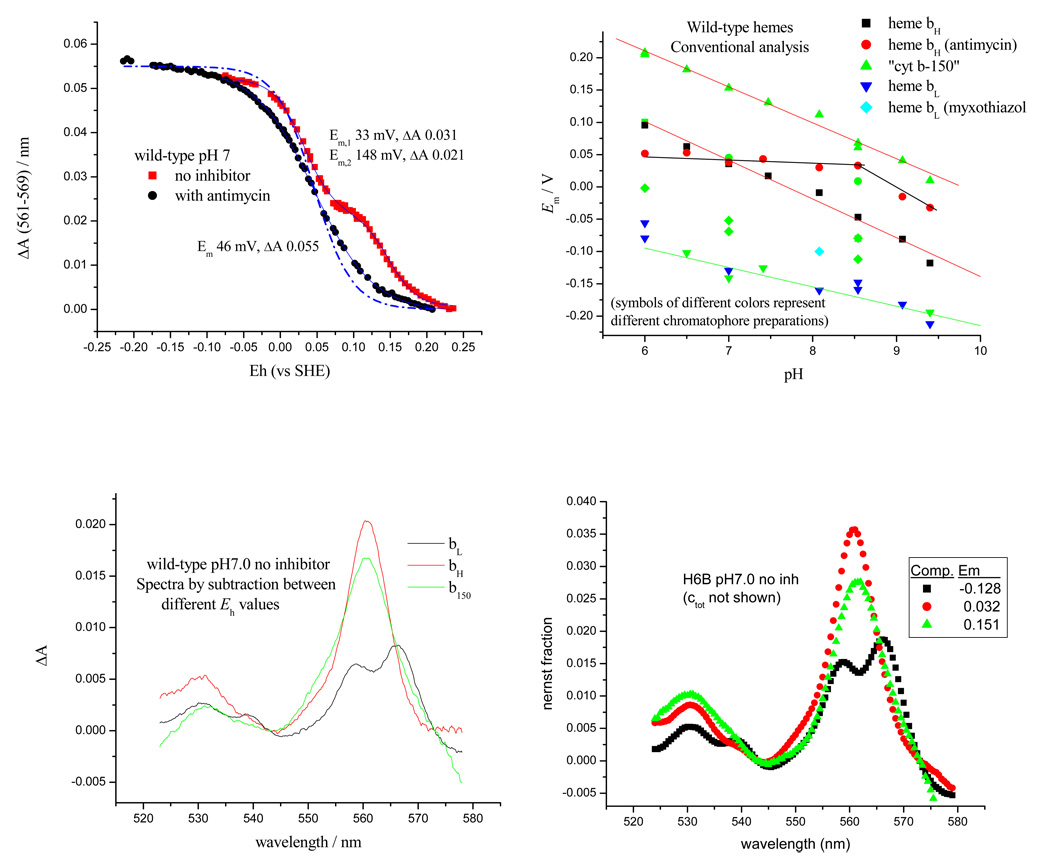
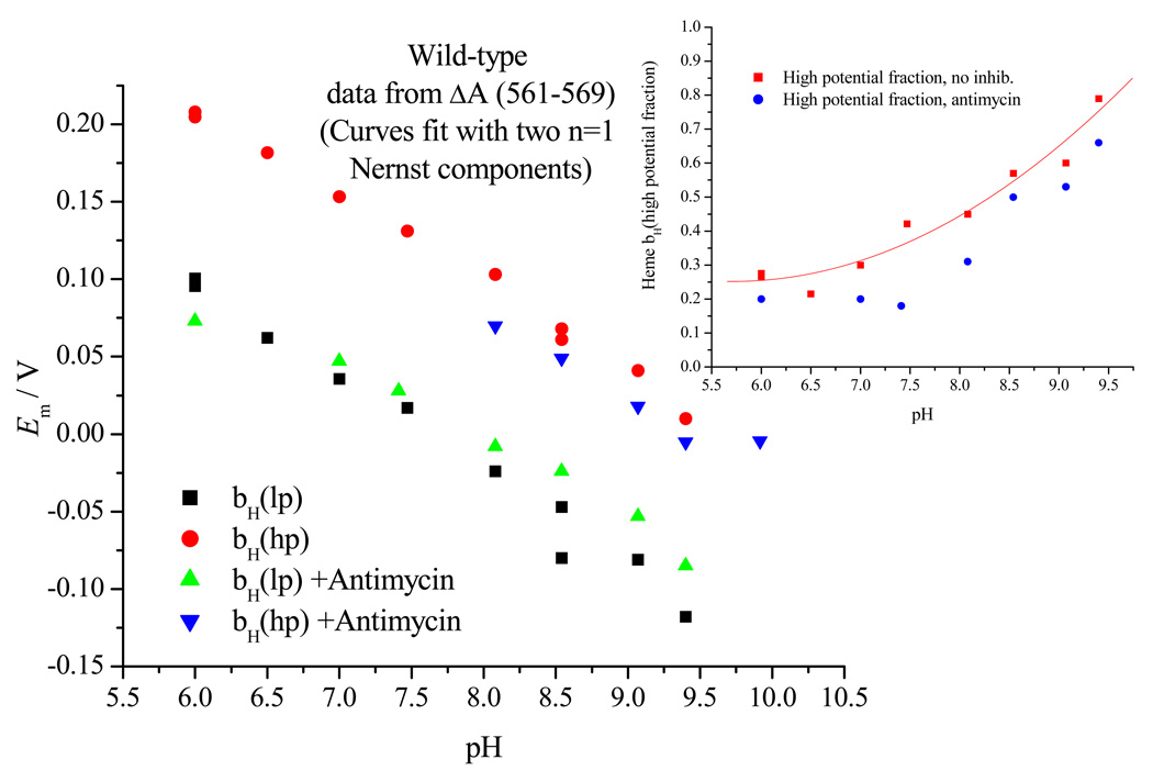
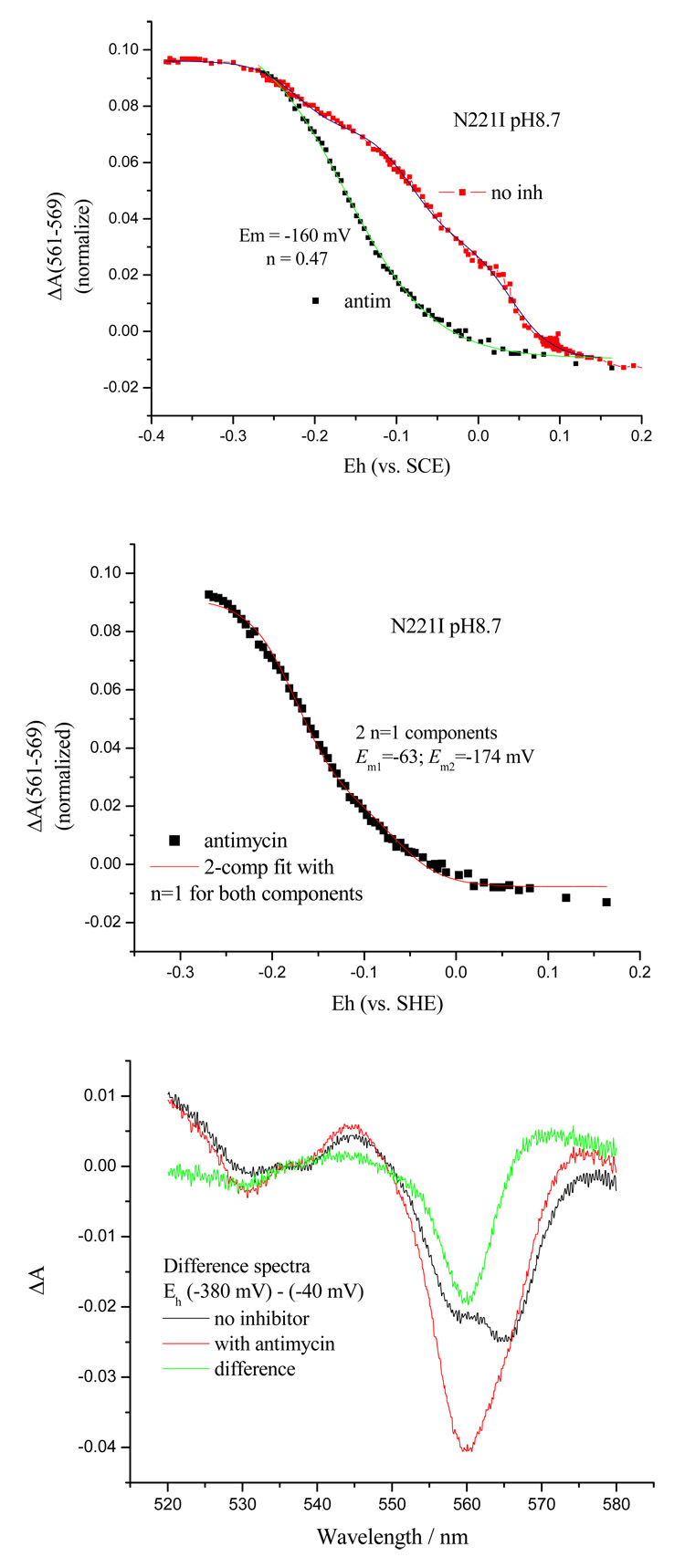
Recent progress in understanding the Q-cycle mechanism of the bc(1) complex is reviewed. The data strongly support a mechanism in which the Q(o)-site operates through a reaction in which the first electron transfer from ubiquinol to the oxidized iron-sulfur protein is the rate-determining step for the overall process. The reaction involves a proton-coupled electron transfer down a hydrogen bond between the ubiquinol and a histidine ligand of the [2Fe-2S] cluster, in which the unfavorable protonic configuration contributes a substantial part of the activation barrier. The reaction is endergonic, and the products are an unstable ubisemiquinone at the Q(o)-site, and the reduced iron-sulfur protein, the extrinsic mobile domain of which is now free to dissociate and move away from the site to deliver an electron to cyt c(1) and liberate the H(+). When oxidation of the semiquinone is prevented, it participates in bypass reactions, including superoxide generation if O(2) is available. When the b-heme chain is available as an acceptor, the semiquinone is oxidized in a process in which the proton is passed to the glutamate of the conserved -PEWY- sequence, and the semiquinone anion passes its electron to heme b(L) to form the product ubiquinone. The rate is rapid compared to the limiting reaction, and would require movement of the semiquinone closer to heme b(L) to enhance the rate constant. The acceptor reactions at the Q(i)-site are still controversial, but likely involve a "two-electron gate" in which a stable semiquinone stores an electron. Possible mechanisms to explain the cyt b(150) phenomenon are discussed, and the information from pulsed-EPR studies about the structure of the intermediate state is reviewed. The mechanism discussed is applicable to a monomeric bc(1) complex. We discuss evidence in the literature that has been interpreted as shown that the dimeric structure participates in a more complicated mechanism involving electron transfer across the dimer interface. We show from myxothiazol titrations and mutational analysis of Tyr-199, which is at the interface between monomers, that no such inter-monomer electron transfer is detected at the level of the b(L) hemes. We show from analysis of strains with mutations at Asn-221 that there are coulombic interactions between the b-hemes in a monomer. The data can also be interpreted as showing similar coulombic interaction across the dimer interface, and we discuss mechanistic implications.


Figures

Fig. 1
Fig. 1. The modified Q-cycle mechanism
The mechanism demonstrated in the 1980’s, superimposed on the Rb. sphaeroides structure (PDB file 2QJK). Electron transfers are shown by blue arrows; H+ release or uptake by red arrows, exchange of Q or QH2 by open blue-green (Qo-site) or yellow (Qi-site) arrows; inhibition sites by cyan arrows. See text for description of operation.
Fig. 2
Fig. 2. Synergy of spectroscopy, crystallography, and thermodynamic analysis
(Left) Structures of wild-type, S154A and Y156F mutants (modified residues shown with electron densities); (middle) 2D ESEEM plots showing proton profiles in wild-type and S154A, and difference (bottom), showing loss of protons a, b (similar data have been obtained for the Y156F mutant); (right) RR spectra showing differences in ISP mutants S154A and Y156F, compared to SDX mutants and wild-type, and (bottom) PFV titrations, showing voltammogram (left, WT, pH 7), and Em vs. pH curves from titration data for wild-type and five mutant strains (right).
Fig. 3
Fig. 3. Schemes to show potential electron transfer pathways involved in electron equilibration between monomers at the level of heme bL
A. The redox centers of the dimer are shown, and the Qo- and Qi-sites indicated by stigmatellin and antimycin, respectively. The scheme shows the dimer with the Qi-site blocked in both monomers by antimycin (blue crosses), and the Qo-site of one monomer blocked by myxothiazol (red cross). Under these conditions, if electron transfer between hemes bL could occur, both hemes bH could be reduced by turnover of the uninhibited Qo-site (broken red arrows). B. The dimer interface, showing residues referred to in the text. The dotted line shows the 10.21 Å distance between heme bL 4-vinyl groups (taken from PDB file 2qjy, chains A–F).
Fig. 4
Fig. 4. Titrations of Qo-site with myxothiazol showing linear curves in WT and mutant strains
a) Typical curves showing the kinetics of heme bH reduction in WT in the presence of antimycin. Similar sets of data were taken for each strain, all of which had similar maximal rates. Myxothiazol concentrations are indicated to the right of each curve. b) Fraction of heme bH reduced at 20 ms (expressed as %) in the presence of Qi-site inhibitor antimycin A, as a function of inhibitor concentration. The system was excited with a xenon flash (~5 µs at half-height) and the reduction of bH monitored from the difference kinetics measured at 561–569 nm. Ambient redox potential was poised at ~100 mV (QH2 oxidation close to maximal). Strains are indicated by labels; bc1 complex in the range 70–220 nM.
Fig. 5
Fig. 5. Coulombic free-energy change expected between hemes
The change in Em expected from coulombic effects between hemes calculated from distance apart, for different intervening dielectric values.
Fig. 5
Fig. 5. Coulombic free-energy change expected between hemes
The change in Em expected from coulombic effects between hemes calculated from distance apart, for different intervening dielectric values.
Fig. 6
Fig. 6. Thermodynamic behavior of the b-type hemes in H6B strain (his-tagged cyt b with wild-type sequence) in situ
(Top left) typical titration data in the absence and presence of antimycin (pH 7); (bottom left) spectra from redox cuts showing cyt b150, heme bH and heme bL; the dashed line shows the curve expected for an n=1 component, with amplitude for degree of reduction plotted as ΔA=0.055110(EhEm)/59+1 (Bottom, right) spectra derived by fitting titrations at each wavelength using fixed values for Em, and n, for each component; (top, right) the pH-dependence of Em values for individual components, assuming single species for each component.
Fig. 7
Fig. 7. Reanalysis of the thermodynamic behavior assuming the coulombic effects are in play
Titration data for heme bH in the absence or presence of antimycin were assumed to reflect two components split by coulombic interaction.
Fig. 8
Fig. 8. Titration of heme bH in chromatophores from N221I in the presence and absence of antimycin
A (top), titration curves showing shift in Em on addition of antimycin; B (middle), titration with antimycin fit by 2-component, n=1; C (bottom), absorbance spectra showing the difference (−380)-(−40) mV from pH 8.7 titration. The difference with and without antimycin shows the contribution of heme bH with its low potential in the presence of antimycin.
Fig. 9
Fig. 9. Scheme to show some of the coulombic interactions suggested
The dimer is represented by four Q-sites (circles) with antimycin blocking Qi (blue cross). The hemes are shown as horizontal bars, either empty, or occupied by an electron (red dot). Top, in wild-type, heme bH has Em,7 ~50 mV, heme bL has in intrinsic Em,7 ~−30 mV, but titrates with Em ~−100 mV because of coulombic interaction with heme bH. Bottom, in N221I, both hemes have the same intrinsic Em,7 ~−30. The first electron can go to either, and change the Em of the other. For clarity only the coulombic interactions within a monomer are shown However, the titrations suggest that additional interactions across the dimer interface may also occur.

References

    1. Osyczka A, Moser CC, Dutton PL. Fixing the Q-cycle. Trends in Biochemical Science. 2005;30:176–182. - PubMed
    1. Crofts AR. The cytochrome bc1 complex – function in the context of structure. Annu. Rev. Physiol. 2004;vol. 66:689–733. - PubMed
    1. Crofts AR. The bc1 complex: what is there left to argue about? In: Wikström M, editor. Biophysical and Structural Aspects of Bioenergetics. Cambridge: Royal Society of Chemistry Publishing; 2005. pp. 123–155.
    1. Crofts AR. Proton-coupled electron transfer at the Qo-site of the bc1 complex controls the rate of ubihydroquinone oxidation. Biochim. Biophys. Acta. 2004;1655:77–92. - PubMed
    1. Crofts AR, Lhee S, Crofts SB, Cheng J, Rose S. Proton pumping in the bc1 complex: A new gating mechanism that prevents short circuits. Biochim. Biophys. Acta. 2006;1757:1019–1034. - PubMed

Publication types

Substances

LinkOut - more resources