

Enhanced surgical imaging: laparoscopic vessel identification and assessment of tissue oxygenation
- PMID: 18501814
- PMCID: PMC2749562
- DOI: 10.1016/j.jamcollsurg.2008.01.013
Enhanced surgical imaging: laparoscopic vessel identification and assessment of tissue oxygenation
Abstract
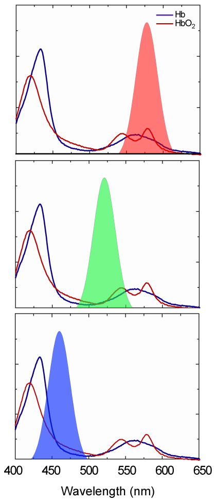
Background: Inherent to minimally invasive procedures are loss of tactile feedback and loss of three-dimensional assessment. Tasks such as vessel identification and dissection are not trivial for the inexperienced laparoscopic surgeon. Advanced surgical imaging, such as 3-charge-coupled device (3-CCD) image enhancement, can be used to assist with these more challenging tasks and, in addition, offers a method to noninvasively monitor tissue oxygenation during operations.
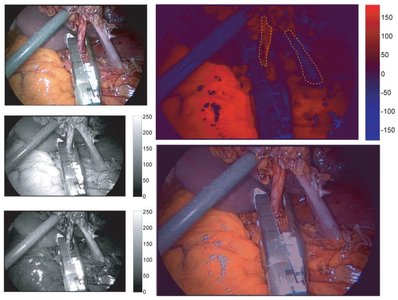
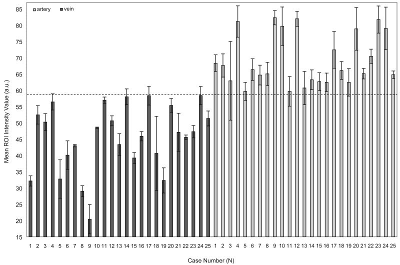
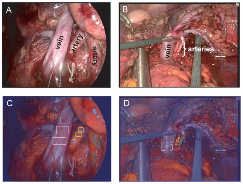
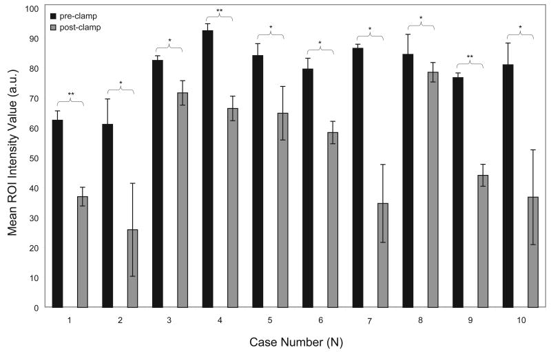
Study design: In this study, 3-CCD image enhancement is used for identification of vessels in 25 laparoscopic donor and partial nephrectomy patients. The algorithm is then applied to two laparoscopic nephrectomy patients involving multiple renal arteries. We also use the 3-CCD camera to qualitatively monitor renal parenchymal oxygenation during 10 laparoscopic donor nephrectomies (LDNs).
Results: The mean region of interest (ROI) intensity values obtained for the renal artery and vein (68.40 +/- 8.44 and 45.96 +/- 8.65, respectively) are used to calculate a threshold intensity value (59.00) that allows for objective vessel differentiation. In addition, we examined the renal parenchyma during LDNs. Mean ROI intensity values were calculated for the renal parenchyma at two distinct time points: before vessel stapling (nonischemic) and just before extraction from the abdomen (ischemic). The nonischemic mean ROI intensity values are statistically different from the ischemic mean ROI intensity values (p < 0.05), even with short ischemia times.
Conclusions: We have developed a technique, 3-CCD image enhancement, for identification of vasculature and monitoring of parenchymal oxygenation. This technique requires no additional laparoscopic operating room equipment and has real-time video capability.
Figures
References
-
- Khauli RB, El-Hout Y, Hussein M, et al. A controlled sequential evaluation of laparoscopic donor nephrectomy versus open donor nephrectomy: an update. Transplant Proc. 2005;37:633–634. - PubMed
-
- Lind MY, Ijzermans JNM, Bonjer HJ. Open vs laparoscopic donor nephrectomy in renal transplantation. Br J Urol Int. 2002;89:162–168. - PubMed
-
- Bykowski J, Kollias N, LaMuraglia GM. Evaluation of peripheral arterial occlusive disease and postsurgical viability using reflectance spectroscopy of skin. Microvasc Res. 2004;67:152–158. - PubMed
-
- Knoefel WT, Kollias N, Rattner DW, Nishioka NS, Warshaw AL. Reflectance soectroscopy of pancreatic microcirculation. J Appl Physiol. 1996;80:116–123. - PubMed
-
- Kostenich G, Kimel S, Peled S, Orenstein A. Monitoring PDT-induced damage using spectrally resolved reflectance imaging of tissue oxygenation. Cancer Letters. 2005;219:169–175. - PubMed
Publication types
MeSH terms
Substances
Grants and funding
LinkOut - more resources
Full Text Sources
