. 2008 Jul 18;283(29):20299-308.
doi: 10.1074/jbc.M800019200. Epub 2008 May 23.

Kinetic analysis of human enzyme RDH10 defines the characteristics of a physiologically relevant retinol dehydrogenase

Affiliations

Kinetic analysis of human enzyme RDH10 defines the characteristics of a physiologically relevant retinol dehydrogenase

Olga V Belyaeva et al. J Biol Chem. .

Abstract

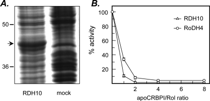
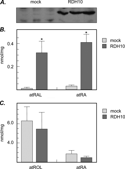
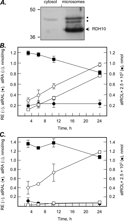
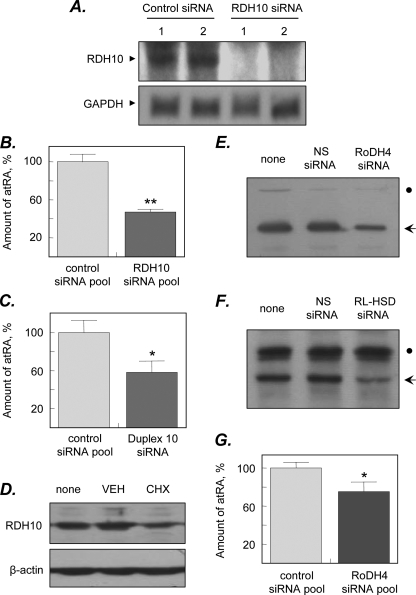
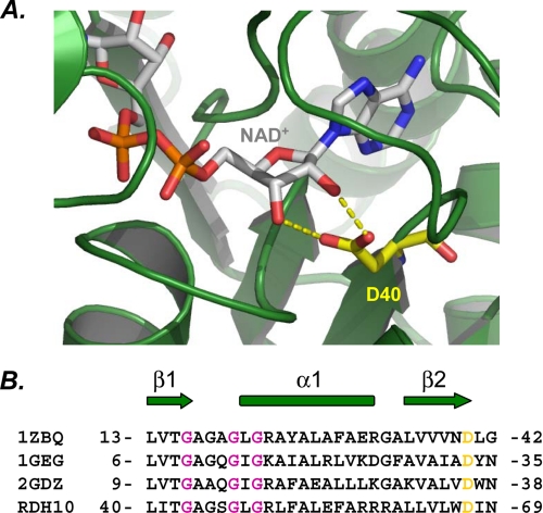
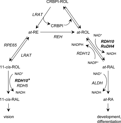
Human retinol dehydrogenase 10 (RDH10) was implicated in the oxidation of all-trans-retinol for biosynthesis of all-trans-retinoic acid, however, initial assays suggested that RDH10 prefers NADP(+) as a cofactor, undermining its role as an oxidative enzyme. Here, we present evidence that RDH10 is, in fact, a strictly NAD(+)-dependent enzyme with multisubstrate specificity that recognizes cis-retinols as well as all-trans-retinol as substrates. RDH10 has a relatively high apparent K(m) value for NAD(+) (~100 microm) but the lowest apparent K(m) value for all-trans-retinol (~0.035 microm) among all NAD(+)-dependent retinoid oxidoreductases. Due to its high affinity for all-trans-retinol, RDH10 exhibits a greater rate of retinol oxidation in the presence of cellular retinol-binding protein type I (CRBPI) than human microsomal RoDH4, but like RoDH4, RDH10 does not recognize retinol bound to CRBPI as a substrate. Consistent with its preference for NAD(+), RDH10 functions exclusively in the oxidative direction in the cells, increasing the levels of retinaldehyde and retinoic acid. Targeted small interfering RNA-mediated silencing of endogenous RDH10 or RoDH4 expression in human cells results in a significant decrease in retinoic acid production from retinol, identifying both human enzymes as physiologically relevant retinol dehydrogenases. The dual cis/trans substrate specificity suggests a dual physiological role for RDH10: in the biosynthesis of 11-cis-retinaldehyde for vision as well as the biosynthesis of all-trans-retinoic acid for differentiation and development.


Figures

FIGURE 1.
FIGURE 1.
Expression of RDH10 in Sf9 cells and effect of apoCRBPI on RDH10 retinol dehydrogenase activity. A, Sf9 cells were infected with recombinant (RDH10-His6) or wild-type (mock) Baculovirus vector and microsomes were isolated by differential centrifugation 3 days later. Microsomal proteins were separated by SDS-PAGE and the proteins were visualized by staining with Coomassie R-250. The position of RDH10 protein is indicated by an arrow. B, the reactions were carried out with microsomal preparation of RDH10 expressed in Sf9 cells (5 μg) in the presence of 1 mm NAD+ for 15 min at 37 °C. RoDH4 expressed in microsomes of Sf9 cells (5 μg) was used for comparison. The concentration of all-trans-retinol (Rol) was 1μm, whereas the concentration of apoCRBPI was varied from 0 to 8 μm. The results shown are mean ± S.D. of three independent experiments and were reproduced with three different preparations of apoCRBPI.
FIGURE 2.
FIGURE 2.
Increase in retinoic acid biosynthesis in RDH10-transfected cells. A, HEK293 cells were transfected with HA-RDH10/pCMV-HA vector (RDH10) or empty vector (mock). Expression of RDH10 was confirmed by Western blotting using HA antibodies. Cells were incubated with all-trans-retinol (2 μm) for 6 h (B) or with all-trans-retinaldehyde (5 μm) for 4 h (C). Retinoid metabolites were analyzed by normal phase HPLC. atROL, all-trans-retinol; atRAL, all-trans-retinaldehyde; atRA, all-trans-retinoic acid. Data are mean ± S.D. (vertical bars), n = 3. Statistical analysis was done using two-tailed unpaired Student's t test comparing RDH12-transfected cells to mock-transfected cells; *, p < 0.001. The results are representative of three independent experiments.
FIGURE 3.
FIGURE 3.
Time course of retinol metabolism in HEK293 cells. A, HEK293 cells were transfected with untagged RDH10 expression construct in pCMV-Tag4a and the microsomes and cytosol were isolated by differential centrifugation. RDH10 protein was detected by Western blotting using RDH10 rabbit polyclonal antiserum. The slower moving bands indicated by dots are due to nonspecific binding of antibodies and are not glycosylated forms of RDH10, because RDH10 polypeptide sequence does not contain any consensus N-glycosylation sites (NxS/T). B, RDH10-transfected cells were incubated with all-trans-retinol (2 μm) for the indicated periods of time. C, cells transfected with empty vector served as controls for endogenous cellular activities. Retinoids were extracted and analyzed by normal phase HPLC as described under “Experimental Procedures.” RE, retinyl esters; other abbreviations are as described in the legend to Fig. 2.
FIGURE 4.
FIGURE 4.
Effect of siRNA-mediated knockdown of SDRs on retinoic acid biosynthesis. A, HEK293 cells were transfected with ON-TARGETplus pool of four siRNAs targeted against RDH10 or siCONTROL Non-targeting siRNA pool. Knockdown of RDH10 mRNA expression (RDH10) was confirmed by Northern blot analysis. A cDNA probe for glyceraldehyde-3-phosphate dehydrogenase (GAPDH) was used as a loading control. Shown are the results of two parallel experiments (1 and 2). B, cells were incubated with 10 μm all-trans-retinol for 24 h and all-trans-retinoic acid (atRA) levels were analyzed by HPLC. Data shown are average ± S.D. of triplicates from one of three experiments; **, p = 0.0004. C, HEK293 cells were transfected with RDH10 duplex 10 (J-01008-10, Dharmacon) or siCONTROL. Non-targeting siRNA pool and the levels of retinoic acid were determined by normal phase HPLC. Data shown are average ± S.D. of triplicates from one of two experiments; *, p = 0.015. D, untagged RDH10 was expressed in HEK293 cells and the cells were treated with either vehicle (VEH) or cycloheximide (CHX) for 36 h. The levels of RDH10 protein in the cells before (none) and after treatments were determined by immunoblotting with RDH10 polyclonal antiserum. Staining with β-actin antibodies was used as a control for loading of samples. E and F, HEK293 cells stably expressing RoDH4 (E) or RL-HSD (F) protein were transfected with the siGENOME SMARTpools M-008311-00 and M-009280-00 (Dharmacon), respectively, and the levels of corresponding proteins were determined by Western blot analysis 3 days after transfections. The positions of RoDH4 and RL-HSD are indicated by arrows. Dots indicate nonspecific staining, which attests to equal loading of the samples. G, cells transfected with RoDH4-specific siRNA pool or control siRNA were incubated with 10 μm retinol for 24 h and the levels of retinoids were analyzed by HPLC as described in B. Data shown are average ± S.D. of triplicates from one of three experiments; *, p = 0.02.
FIGURE 5.
FIGURE 5.
Cofactor specificity of RDH10. A, ribbon representation of the dehydrogenase domain of human 17β-HSD4 (PDB code 1ZBQ). Shown is the view of the cofactor binding region of the Rossmann fold with bound NAD+. NAD+ and D40 are shown as stick models (CPK, carbon gray and CPK, carbon yellow, respectively). Hydrogen bonds between Asp-40 and NAD+ are shown in yellow. Molecular graphics for structural representation were made using PyMol (DeLano Scientific LLC, Palo Alto, CA). B, structural alignment of NAD+-dependent SDRs. The secondary structure elements are indicated as arrows for β-strands and as cylinders for α-helices. Conserved aspartate residues that determine the preference for NAD+ are highlighted in yellow. Gly residues that constitute the GXXXGXG cofactor-binding motif are shown in dark pink. 1GEG, crystal structure of meso-2,3-butanediol dehydrogenase in a complex with NAD+ and inhibitor mercaptoethanol; 2GDZ, crystal structure of 15-hydroxyprostaglandin dehydrogenase type 1, complexed with NAD+.
FIGURE 6.
FIGURE 6.
Schematic of retinoid metabolic pathways. The schematic illustrates the role of RDH10 in the biosynthesis of all-trans-retinoic acid for development and differentiation and in the biosynthesis of 11-cis-retinaldehyde for vision. The 11-cis-retinol dehydrogenase activity of RDH10 is marked with a star to indicate that this activity has not yet been demonstrated in vivo. REH, retinyl ester hydrolase; RPE65, isomerohydrolase; ALDH, aldehyde dehydrogenase. Other abbreviations are as described in the legend to Fig. 2. Note that the NAD+-dependent RDH10 and RoDH4 function in the oxidative direction, whereas the NADP+-dependent RDH12 functions in the reductive direction. Lecithin:retinol acyltransferase (LRAT) appears to utilize all-trans-retinol bound to CRBPI, whereas the oxidoreductases exclusively recognize the unbound all-trans-retinol as a substrate.

References

    1. Mangelsdorf, D., Umesono, K., and Evans, R. M. (1994) in The Retinoids: Biology, Chemistry and Medicine (Sporn, M. B., Roberts, A. B., and Goodman, D. S., eds) pp. 319-350, Raven Press, New York
    1. Napoli, J. L. (1999) Biochim. Biophys. Acta 1440 139-162 - PubMed
    1. Duester, G. (1996) Biochemistry 35 12221-12227 - PubMed
    1. Kedishvili, N. Y. (2002) Curr. Org. Chem. 6 1247-1257
    1. Veech, R. L., Eggleston, L. V., and Krebs, H. A. (1969) Biochem. J. 115 609-619 - PMC - PubMed

Publication types

MeSH terms

LinkOut - more resources