. 2008 Aug 15;283(33):22628-36.
doi: 10.1074/jbc.M800503200. Epub 2008 May 23.

Structure and mechanism of the rebeccamycin sugar 4'-O-methyltransferase RebM

Affiliations

Structure and mechanism of the rebeccamycin sugar 4'-O-methyltransferase RebM

Shanteri Singh et al. J Biol Chem. .

Abstract

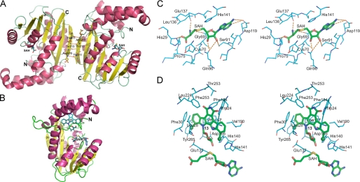
The 2.65-angstroms crystal structure of the rebeccamycin 4'-O-methyltransferase RebM in complex with S-adenosyl-l-homocysteine revealed RebM to adopt a typical S-adenosylmethionine-binding fold of small molecule O-methyltransferases (O-MTases) and display a weak dimerization domain unique to MTases. Using this structure as a basis, the RebM substrate binding model implicated a predominance of nonspecific hydrophobic interactions consistent with the reported ability of RebM to methylate a wide range of indolocarbazole surrogates. This model also illuminated the three putative RebM catalytic residues (His140/141 and Asp166) subsequently found to be highly conserved among sequence-related natural product O-MTases from GC-rich bacteria. Interrogation of these residues via site-directed mutagenesis in RebM demonstrated His140 and Asp166 to be most important for catalysis. This study reveals RebM to be a member of the general acid/base-dependent O-MTases and, as the first crystal structure for a sugar O-MTase, may also present a template toward the future engineering of natural product MTases for combinatorial applications.


Figures

FIGURE 1.
FIGURE 1.
A, naturally occurring indolocarbazoles staurosporine (1), K252a (2), rebeccamycin (3), and AT2433A1 (4). B, the putative biosynthetic pathways for 1, 3, and 4. The reaction catalyzed by RebM is highlighted in the box.
FIGURE 2.
FIGURE 2.
A, quaternary structure of the RebM-AdoHcy (SAH) ternary complex. Residues in the dimer interface are represented as stick models with the orange lines indicating putative hydrogen bonding interactions between the two subunits. B, ribbon diagram of the RebM monomer substrate complex with labeled amino (N) and carboxyl (C) termini and the secondary structure elements. Aglycon (13) and AdoHcy are represented in cyan, and the helices and strands are colored in pink and yellow, respectively. C, stereoview of RebM-AdoHcy interactions. The stick model of AdoHcy is depicted in green, and the interacting RebM residues are labeled and illustrated in cyan. D, stereoview of RebM-13 modeled interactions. AdoHcy and 13 (both green) are positioned in a linear arrangement to facilitate SN2 nucleophilic attack. Interacting RebM residues are labeled and illustrated in cyan.
FIGURE 3.
FIGURE 3.
Comparison of the overall topologies of structural homologs. A, structural overlay of RebM from L. aerocolonigenes (Protein Data Bank code 3BUS) colored pink and sarcosine dimethylglycine methyltransferase from Galdieria sulfuraria (Protein Data Bank code 2O57) colored green (root mean square (RSM) deviation between the structures is 1.8 Å). B, structural overlay of RebM from L. aerocolonigenes (Protein Data Bank code 3BUS) colored pink and mycolic acid synthase Hma (Mmaa4) from Mycobacterium tuberculosis (Protein Data Bank code 2FK8) colored blue (root mean square (RSM) deviation between the structures is 2.0 Å). AdoHcy (SAH) is in stick model and colored yellow. The molecular graphics program PyMOL was used in generating these graphics. C, sequence alignment of RebM from L. aerocolonigenes (Protein Data Bank code 3BUS), sarcosine dimethylglycine methyltransferase (GNMT, glycine N-methyltransferase) from G. sulfuraria (Protein Data Bank code 2O57), mycolic acid synthase Hma (Mmaa4) (Protein Data Bank code 2FK8) from M. tuberculosis, mycolic acid cyclopropane synthase (Cmaa1) (Protein Data Bank code 1KPH) from M. tuberculosis, and carminomycin 4-O-methyltransferase (DnrK) (Protein Data Bank code 1TW3) from Streptomyces peucetius. In this alignment, the predicted secondary structure is illustrated above the sequence. Helices and sheets are colored red and green, respectively, wherein yellow-colored helices α8, α9, and α10 indicate incomplete sequence. Loops involved in cofactor binding are labeled (L1–L3), and sequence of blue color designates the AdoMet binding core domain (β1–β7 and α1–α3, α5, and α7). Highly or moderately conserved residues are colored red and orange, respectively, with putative catalytic residues (based upon RebM mutagenesis) colored green.
FIGURE 4.
FIGURE 4.
Alignment of selected RebM homologs identified in a PSI-BLAST search. Numbers within parentheses are the accession codes from the Swiss Protein Data Bank. Shown from top to bottom are RebM from L. aerocolonigenes (BAC10678.1), AtM from Actinomadura melliaura (ABC02795.1, 56% identity), RifMT (rifamycin O-methyltransferase) from Amycolatopsis mediterranei (AAC01738.1, 53% identity), MonE (monensin 3-O-methyl transferase) from Streptomyces cinnamonensis (AAO65792.1, 40% identity), MitM from Streptomyces lavendulae (AAD28459.1, 43% identity), SnogM from Streptomyces nogalater (AAG42853.1, 40% identity), AveD (C5-O-methyltransferase) from Streptomyces avermitilis MA-4680 (NP_822112.1, 41% identity), NigE from Streptomyces violaceusniger (ABC84455.1, 41% identity), and MitN from S. lavendulae (AAD28458.1, 36% identity). In this alignment, the amino acid numbering refers to RebM, and the secondary structure of RebM is also illustrated (α-helices as red cylinders and β-strands as green arrows). RebM loops involved in cofactor binding are labeled (L1–L3), and RebM secondary structures are labeled β1–β7 and α1–α10. The text of highly or moderately conserved residues are colored red and blue, respectively. Yellow and green highlighted boxes designate regions involved in AdoMet binding and putative catalytic residues (based upon RebM mutagenesis). The program “Multalin version 5.4.1” was used for the multiple sequence alignment using the symbol comparison table blosum62 with a gap weight of 12 and a gap length weight of 2. Consensus symbols include:!, Ile or Val; $, Leu or Met; %, Phe or Tyr; #, Asn, Asp, Gln, or Glu;..., variable sequence in the alignment.

References

    1. Prudhomme, M. (2000) Curr. Med. Chem. 7 1189-1212 - PubMed
    1. Long, B. H., and Balasubramanian, B. N. (2000) Expert Opin. Ther. Pat. 10 635-666
    1. Bailly, C. (2000) Curr. Med. Chem. 7 39-58 - PubMed
    1. Tamaoki, T., Nomoto, H., Takahashi, I., Kato, Y., Morimoto, M., and Tomita, F. (1986) Biochem. Biophys. Res. Commun. 135 397-402 - PubMed
    1. Sanchez, C., Mendez, C., and Salas, J. A. (2006) Nat. Prod. Rep. 23 1007-1045 - PubMed

Publication types

MeSH terms

Associated data

LinkOut - more resources