

Review
doi: 10.1182/blood-2008-01-078204.
The vessel wall and its interactions
Affiliations
- PMID: 18502843
- PMCID: PMC2396724
- DOI: 10.1182/blood-2008-01-078204
Item in Clipboard
Review
The vessel wall and its interactions
Blood.
.
Abstract
Blood cell interactions with the vessel wall were first documented almost 170 years ago. Modern advances have revealed that leukocyte and platelet interactions with the endothelium are at the nexus of complex, dynamic cellular and molecular networks that, when dysregulated, may lead to pathological inflammation and thrombosis, which are major sources of morbidity and mortality in the Western world. In this review, we relate the history of blood cell interactions with the vasculature, discuss recent progress, and raise some unresolved questions awaiting the field.
Figures

Drawing by Rudolph Wagner; legend translated from the German. Small venous branch a of the webbing of Rana temporaria at 350× magnification and close to the surface of the epidermis, whose cobblestone-like, mostly hexagonal, flattened, and for the most part nucleated cells b,b,b,b go over the vessel. Blood corpuscles are seen in multiple rows partially on the flat side, partially standing on the edge; in the light area between the flow of blood corpuscles and the vessel wall surrounded by several parallel filaments, one can see the round, bright, much slower moving lymph-corpuscles. The whole image was prepared at low illumination.

Weibel-Palade bodies are endothelial-specific organelles that store von Willebrand factor. (Left) Oblique section of a Weibel-Palade body from pulmonary artery endothelium of a rat, showing parallel arrangement of internal tubules (H). Reproduced with permission from Weibel and Palade. (Right) Electron micrograph (Wagner et al) of human umbilical vein endothelial cells stained for von Willebrand factor with peroxidase showing a cluster of positively stained Weibel-Palade bodies. Left bar is 0.1 μM, and right bar is 0.5 μM. Reproduced from The Journal of Cell Biology., Copyright 1964 and 1982 The Rockefeller University Press.
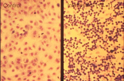
Neutrophils adhere to cytokine-stimulated cultured endothelial cells. Phase-contrast photomicrograph of the adhesion of human neutrophils to control (left) and interleukin-1–treated (right) human umbilical vein endothelial monolayers taken at the end of a 10-minute adhesion assay. Reproduced with permission from American Society for Clinical Investigation.

View of human aorta stained with Sudan III for fat deposits/atheroma (dark stains) from the intimal side. Outer wall branches (experiencing disturbed flow) of the celiac, superior mesenteric, and renal arteries show more extensive staining than the inner walls where shear rates are high and laminar. Reproduced with permission from Macmillan Publishers Ltd.

Multiple sequential steps mediating leukocyte recruitment during inflammation. Leukocytes are captured and begin to roll on P- and E-selectins and their ligands P-selectin glycoprotein ligand-1 (PSGL-1) and E-selectin ligand-1 (ESL-1). Some leukocytes such as lymphocytes or hematopoietic stem and progenitor cells also roll on α4 integrin and its endothelial receptor vascular cell adhesion molecule-1 (VCAM-1). L-selectin is critical for lymphocyte rolling on HEVs in lymphoid tissues. As inflammation progresses, leukocyte rolling velocity decreases, allowing the integration of activation signals from selectin ligands and G-protein–coupled receptors (GPCRs). These activation signals lead to the polarization of slowly rolling leukocytes and clustering of L-selectin and PSGL-1 to a major pole that allows further leukocyte recruitment through secondary tethers via leukocyte-leukocyte interactions. Leukocyte activation enhances integrin affinity and avidity, leading to firm adhesion on intercellular adhesion molecule-1 (ICAM-1) expressed on endothelial cells. Adherent leukocytes continuously migrate laterally to survey the microvasculature and search for possible sites for transmigration. Leukocytes can transmigrate classically through the junctional (paracellular) pathways via interactions among junctional adhesion molecules (JAMs), CD99 and platelet/endothelial-cell adhesion molecule-1 (PECAM-1), endothelial cell–selective adhesion molecule (ESAM), or alternatively through the endothelial cell (transcellular pathway). Illustration by Marie Dauenheimer.

Polarization of adherent leukocytes. In vivo imaging using high-speed high-resolution multichannel fluorescence intravital videomicroscopy of αLβ2 integrin (blue), PSGL-1 (red), and the granulocyte marker Gr-1 (green) expressed on the leukocyte surface in tumor necrosis factor-α (TNF-α) inflamed venules. The majority of leukocytes migrates laterally in inflamed venules. These migrating leukocytes exhibit marked clustering of PSGL-1 at the trailing edge. The large PSGL-1 cluster may contribute to leukocyte recruitment through leukocyte-leukocyte secondary tethers. In contrast, the expression of αLβ2 is relatively homogenous on the leukocyte surface. Reproduced with permission from Nature Publishing Group.

Phase-contrast intravital microscopy showing a platelet rolling on a mesenteric venule after stimulation by the calcium ionophore A23187. “o” indicates the location of the platelet at time 0. Arrowheads point toward the rolling platelet at 0 seconds (top), 1.58 seconds (middle), and 2.96 seconds (bottom). A single much larger and more slowly rolling leukocyte is also seen. Bar represents 30 μm. Reproduced with permission from PNAS.

Platelets roll/translocate on stimulated endothelium of veins; this can lead to their firm adhesion. Endothelium either activated to release WPBs or inflamed by cytokines captures resting or activated platelets. These roll on the selectins or move laterally on VWF. In inflammation, fibrinogen or fibrin oligomers can promote firm adhesion of activated platelets by cross-linking the major platelet integrin to endothelial receptors. These adherent platelets expressing P-selectin may recruit leukocytes but may also initiate pathological thrombosis such as occurs in deep veins. The various known ligands and receptors participating in these platelet adhesion events are listed. Illustration by Marie Dauenheimer.

Activated platelets propel the vicious circle of inflammation. A procoagulant state or inflammatory process generating tissue factor (TF) may lead to platelet activation. Activated platelets bind to leukocytes promoting, in turn, leukocyte activation. Platelet leukocyte complexes produce chemokines that, when deposited on the vessel wall, facilitate leukocyte recruitment. Binding to leukocytes causes platelet P-selectin shedding. Elevated soluble P-selectin activates additional leukocytes. Activated leukocytes can produce leukocyte-derived microparticles (MPs) that further promote endothelial activation as well as cytokines and reactive oxygen species (ROSs) advancing inflammation and endothelial dysfunction. Endothelial dysfunction reduces NO and prostaglandin I2 production leading to increased release of WPBs promoting leukocyte and platelet rolling and facilitating platelet activation. Illustration by Marie Dauenheimer.


References
-
- Wagner R. Leipzig, Germany: Leopold Voss; 1839. Erlauterungstaflen zur Physiologie und Entwicklungsgeschichte.
-
- Waller A. London, England: Edinb Dubl Phil Mag; 1846. Microscopic examination of some principal tissues of the animal frame as observed in the tongue of the living frog, toad, etc. Vol 29. pp. 271–297.
-
- Cohnheim J. London, England: The New Sydenham Society; 1889. Lectures on General Pathology: A Handbook for Practitioners and Students.
-
- Metchnikoff E. London, England: Paul Kegan, Trubner, Trench; 1893. Lectures on the comparative pathology of inflammation. Vol xii.
-
- Jackson SP. The growing complexity of platelet aggregation. Blood. 2007;109:5087–5095. - PubMed
Publication types
MeSH terms
Grants and funding
LinkOut - more resources
Full Text Sources
Other Literature Sources
