. 2008 Sep;49(9):4026-35.
doi: 10.1167/iovs.08-1807. Epub 2008 May 23.

HVEM and nectin-1 are the major mediators of herpes simplex virus 1 (HSV-1) entry into human conjunctival epithelium

Affiliations

HVEM and nectin-1 are the major mediators of herpes simplex virus 1 (HSV-1) entry into human conjunctival epithelium

Jihan Akhtar et al. Invest Ophthalmol Vis Sci. 2008 Sep.

Abstract

Purpose: The human conjunctiva is a natural target for herpes simplex virus (HSV)-1 infection. The goals of this study were to investigate the cellular and molecular mechanisms of HSV-1 entry into human conjunctival epithelial (HCjE) cells. Specific features of entry studied included the method of initial viral binding to cells, pH dependency, and expression and usage of specific HSV-1 entry receptors.

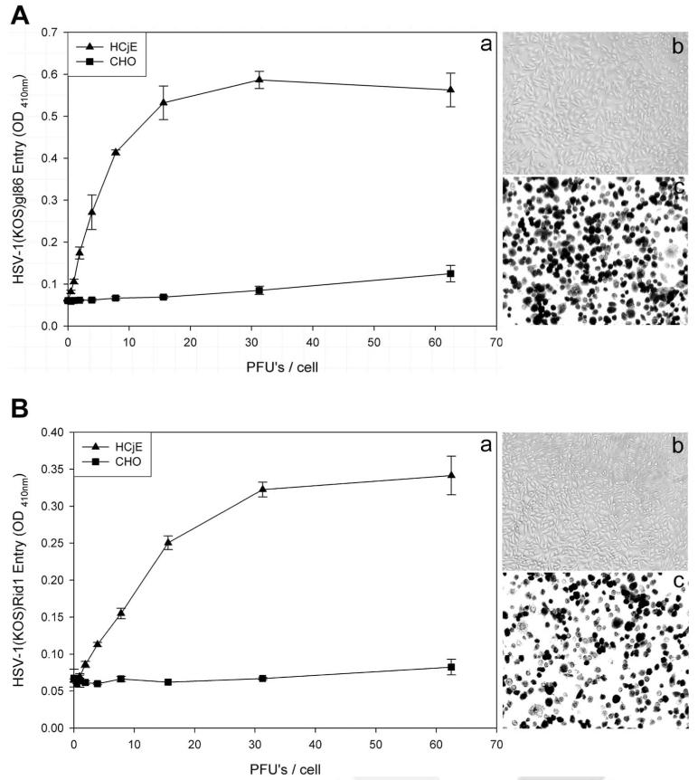
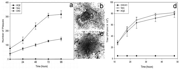
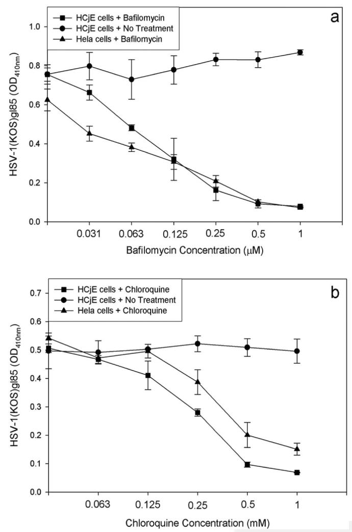
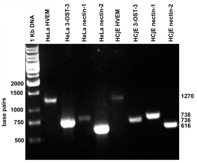
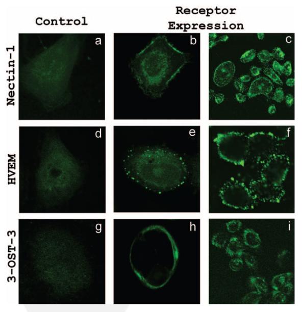
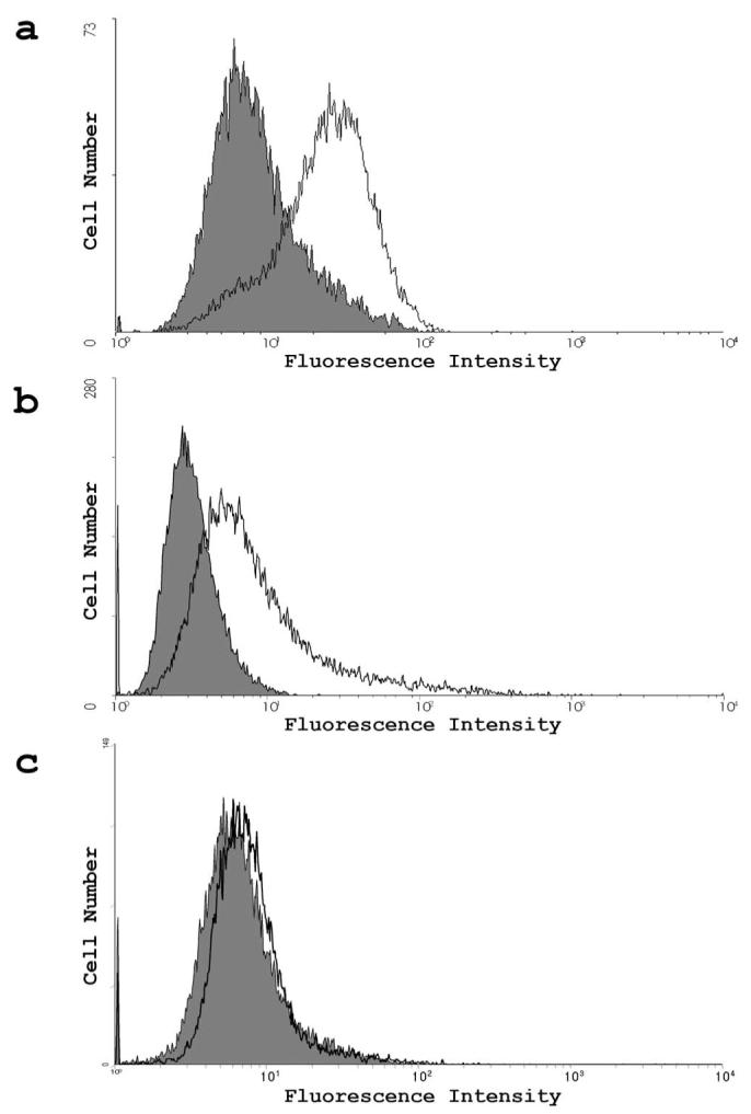
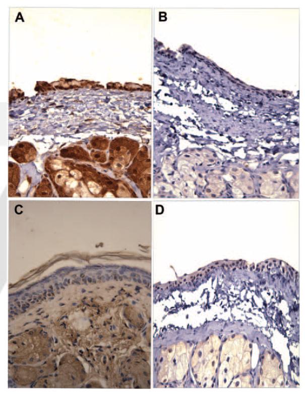
Methods: To observe HSV-1 initial binding, live cell imaging was performed on HSV-1-infected HCjE cells. Reporter HSV-1 virions expressing beta-galactosidase were used to determine entry of wild-type HSV-1(KOS) and a mutant, HSV-1(KOS)Rid1, into HCjE cells. HSV-1 replication in HCjE cells was determined by plaque assays. Lysosomotropic agents were used to determine whether viral entry was pH dependent. Reverse transcription (RT)-PCR, flow cytometry, and immunohistochemistry were used to determine the expression of receptors. Receptor-specific siRNAs were used to define the role of individual entry receptors.

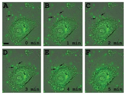
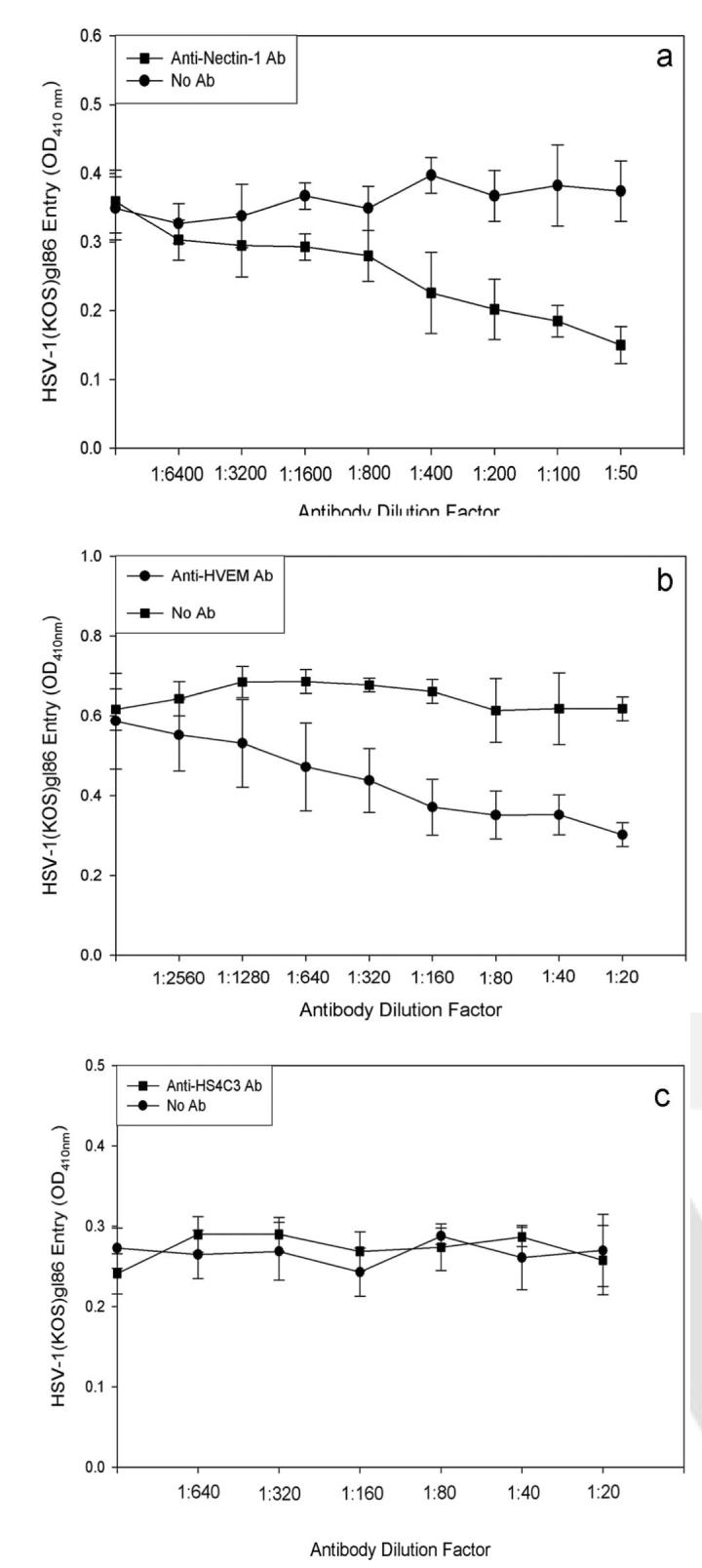
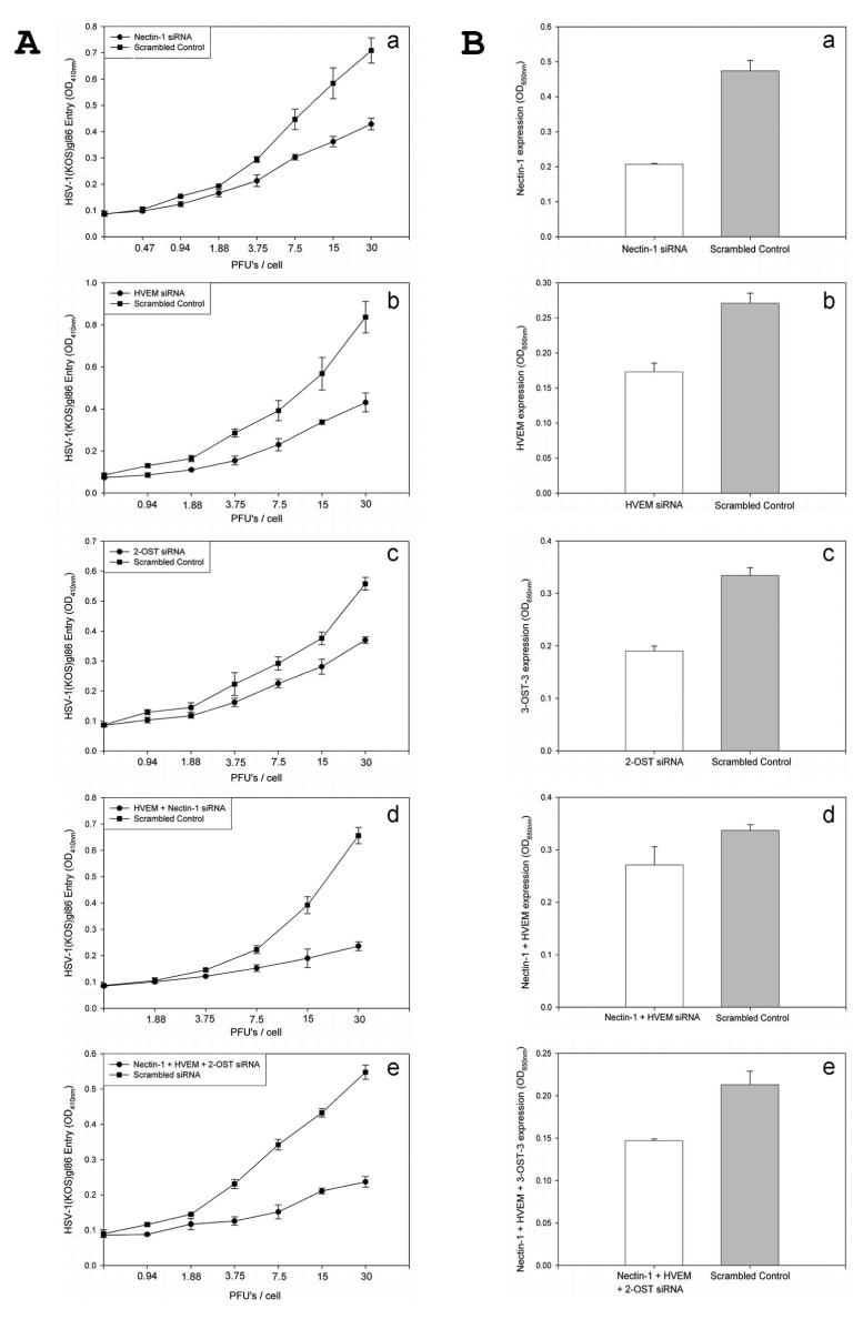
Results: HSV-1 virions attach to filopodia present on HCjE cells and use them to reach the cell body for entry. Cultured HCjE cells demonstrate susceptibility to HSV-1 entry and form plaques confirming viral replication. Blocking vesicular acidification significantly reduces entry, implicating a pH-dependent mode of entry. Multiple assays confirm the expression of entry receptors nectin-1, HVEM, and 3-O-sulfated heparan sulfate (3-OS HS) on the HCjE cell membrane. Knocking down of gD receptors by siRNAs interference implicates nectin-1 and HVEM as the major mediators of entry.

Conclusions: HSV-1 entry into HCjE cells is a pH-dependent process that is aided by targeted virus travel on filopodia. HCjE cells express all three major entry receptors, with nectin-1 and HVEM playing the predominant role in mediating entry.


Figures

FIGURE 1
FIGURE 1
HSV-1 travels along filo-podia on HCjE cells. (AF) Time course of green fluorescence protein expressing HSV-1 (K26GFP) interaction with filopodia. Virions (MOI, 200) were added to HCjE cells and monitored under a fluorescence microscope. Time 0 (A) is when virus (green) is first detected on filopodial surface (arrow). Scale bar, 10 μm.
FIGURE 2
FIGURE 2
Wild-type and a mutant form of HSV-1(KOS) can enter into HCjE cells. (A) Analysis of wild-type HSV-1 entry into HCjE cells. HCjE or control CHO-K1 cells were plated in 96-well culture dishes and inoculated by serial dilutions of β-galactosidase– expressing recombinant HSV-1(KOS) gL86 virus at the plaque-forming unit (PFU)/cell indicated (a). The soluble substrate, ONPG, was added to cells at 6 hours after infection, and the enzymatic activity was measured with a spectrophotometer (a). Similar as-says were repeated with CHO-K1 (b) and HCjE (c) using an insoluble substrate, X-gal, to monitor individual cells turning blue because of virus entry (shown in black). (B) Analysis of HSV-1(KOS) rid1 mutant entry into HCjE cells. HCjE or the control CHO-K1 cells were plated in 96-well culture dishes and inoculated by serial dilutions of β-galactosidase–expressing recombinant HSV-1(KOS)Rid1tk12 virus at the PFU/cell indicated. ONPG (a) and X-gal (b, c) entry assays were repeated, as described.
FIGURE 3
FIGURE 3
HSV-1 can productively replicate in HCjE cells. Confluent monolayers of HCjE, Vero, and wild-type CHO-K1 cells were infected with HSV-1(KOS) at 0.01 PFU/cell for 90 minutes at 37°C. Inoculums were harvested at 24 to 96 hours after infection. Infectious virus titer (PFU/mL) determined in triplicate in Vero cells by plaque assay indicates that the viral titer in cultured HCjE during that time. Data represent the mean ± SD of results in triplicate wells in a representative experiment (a). A typical plaque formed by Vero cells stained with Giemsa stain at 48 hours of infection with HSV-1(KOS) virus is shown (b). A typical Giemsa-stained plaque formed by HCjE cells at 48 hours of infection with HSV-1(KOS) is shown (c). Viral titers, to prove infectivity, were determined on Vero cells (d).
FIGURE 4
FIGURE 4
Lysosomotropic agents negatively affect HSV-1 entry. Monolayers of HCjE cells were either mock treated (No Treatment) or treated with indicated concentrations of bafilomycin (a) or chloroquine (b) and exposed to HSV-1 (50 PFU/cell). HeLa cells were also subjected to the same treatments and served as the positive control. Viral entry was measured using a spectrophotometer 6 hours after infection. Data shown are the means of triplicate measures and are representative of three independent experiments. The control (No Treatment) represents entry into corresponding mock-treated cells.
FIGURE 5
FIGURE 5
Detection of entry receptor mRNA. RT-PCR analysis of the expression of nectin-1, HVEM, and 3-OST-3 (as a surrogate marker for 3-OS HS)–specific messages in HCjE and HeLa cells (served as a control). cDNAs were produced from total RNA isolated from the cells. PCR products were separated by electrophoresis on an agarose gel and stained with ethidium bromide. Molecular weight markers are indicated.
FIGURE 6
FIGURE 6
Immunofluorescence imaging of receptors on HCjE cell membrane. Images shown were taken using the FITC channel of confocal microscope. Cells were blocked for 40 minutes, washed, and either mock treated with buffer alone (a, d, g) or treated with primary antibodies for nectin-1 (b, c), HVEM (e, f), and 3-OS HS(h, i). Images were taken after the incubation of HCjE cells with FITC-conjugated secondary antibodies. Staining of cells with green demonstrated receptor expression.
FIGURE 7
FIGURE 7
Flow cytometry measurement of receptor expression. (a-c) Expression was detected by FACS analysis. Cells were treated with primary antibodies for nectin-1 (a), HVEM (b), and 3-OS HS(c). Controls were treated with secondary antibodies only and are shown in gray on the graph.
FIGURE 8
FIGURE 8
Confirmation of receptor expression by immunohisto-chemistry. Sections of palpebral conjunctiva from adult female BALB/c mice were reacted with anti–nectin-1 (A) or anti–HVEM (C) antibody, and immunohistochemistry was performed as described. Specific staining of the tissues is shown in brown. Note that staining for nectin-1 (A) was relatively stronger than for HVEM (C). Sections stained without primary antibodies were used as controls (B, D).
FIGURE 9
FIGURE 9
Antibody blocking of receptors. Monolayers of HCjE cells were blocked with anti-nectin-1 antibody (a), anti–HVEM antibody (b), and anti–HS4C3 antibody for 2 hours and then were exposed to HSV-1 (50 PFU/cell). Viral entry was measured using a spectrophotometer 6 hours after infection. Data shown are the means of triplicate measures and are representative of three independent experiments. Control (No Ab) represents entry into corresponding mock-treated cells.
FIGURE 10
FIGURE 10
SiRNA downregulation of receptor expression. (A) Viral entry was determined in cells transfected with siRNA against entry receptors: nectin-1 (a), HVEM (b), 2-OST (c), nectin-1 and HVEM (d), and nectin-1, HVEM, and 2-OST (e). Cells transfected with an equal amount of scrambled siRNA were used as control. (B) Determination of surface expression of the receptors by cell-ELISA. Cell-ELISA was performed with cells transfected with siRNA against entry receptors nectin-1 (a), HVEM (b), 2-OST (c), nectin-1 and HVEM (d), and nectin-1, HVEM, and 2-OST. Primary antibodies specific to each receptor and FITC-conjugated secondary antibodies were used for the ELISA. Data shown are the means of triplicate measures.

References

    1. Marangon FB, Miller D, Alfonso E. Laboratory results in ocular viral diseases: implications in clinical-laboratory correlation. Arq Bras Oftalmol. 2007;79:189–194. - PubMed
    1. Liesegang TJ. Herpes simplex virus epidemiology and ocular importance. Cornea. 2001;20:1–13. - PubMed
    1. Uchio E, Takeuchi S, Itoh N, Matsuura N, Ohno S, Aoki K. Clinical and epidemiological features of acute follicular conjunctivitis with special reference to that caused by herpes simplex virus type 1. Br J Ophthalmol. 2000;84:968–972. - PMC - PubMed
    1. Ritterband DC, Friedberg DN. Virus infections of the eye. Rev Med Virol. 1998;8:187–201. - PubMed
    1. Inatomi T, Spurr-Michaud S, Tisdale AS, Gipson IK. Human corneal and conjunctival epithelia express MUC1 mucin. Invest Ophthalmol Vis Sci. 1995;36:1818–1827. - PubMed

Publication types

MeSH terms