. 2008;59(9):2499-512.
doi: 10.1093/jxb/ern114. Epub 2008 May 23.

Truncated myosin XI tail fusions inhibit peroxisome, Golgi, and mitochondrial movement in tobacco leaf epidermal cells: a genetic tool for the next generation

Affiliations

Truncated myosin XI tail fusions inhibit peroxisome, Golgi, and mitochondrial movement in tobacco leaf epidermal cells: a genetic tool for the next generation

Imogen A Sparkes et al. J Exp Bot. 2008.

Abstract

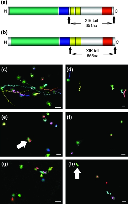
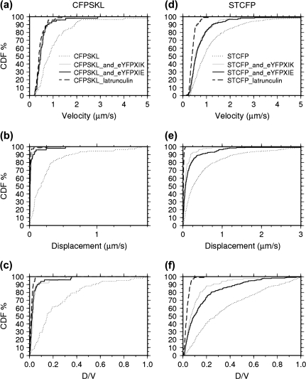
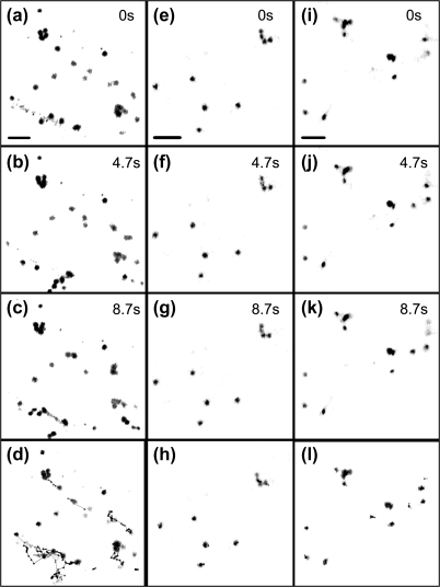
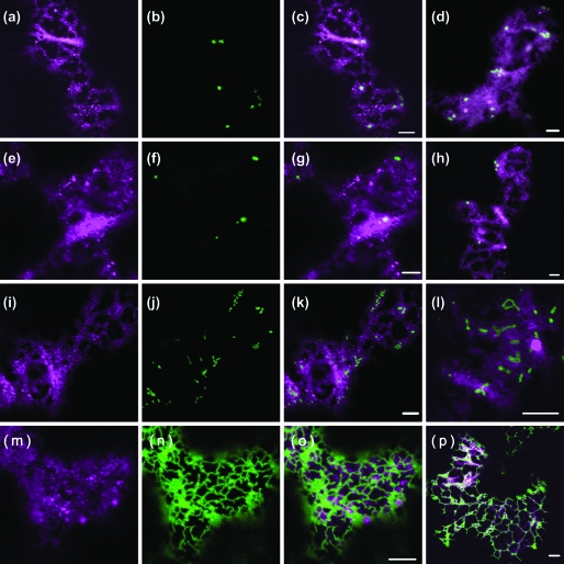
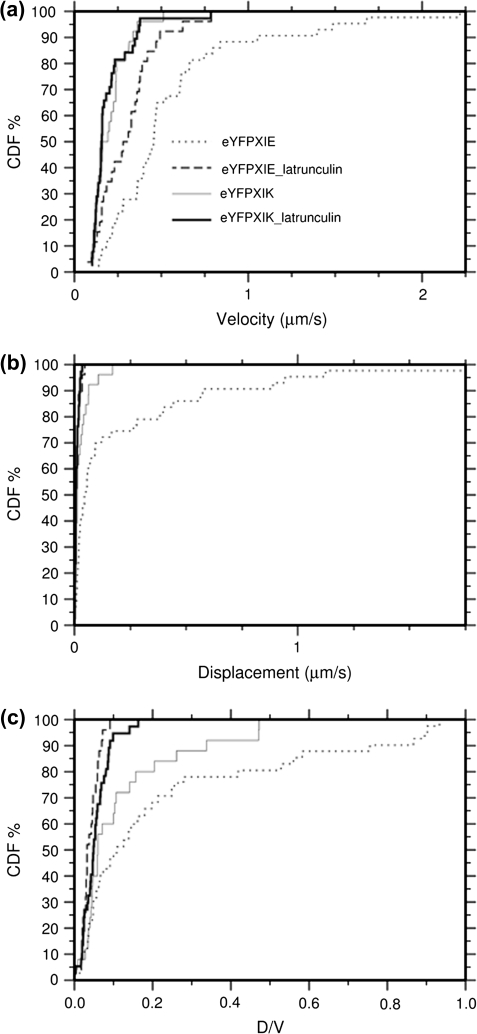
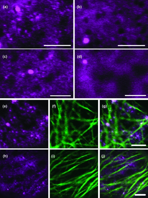
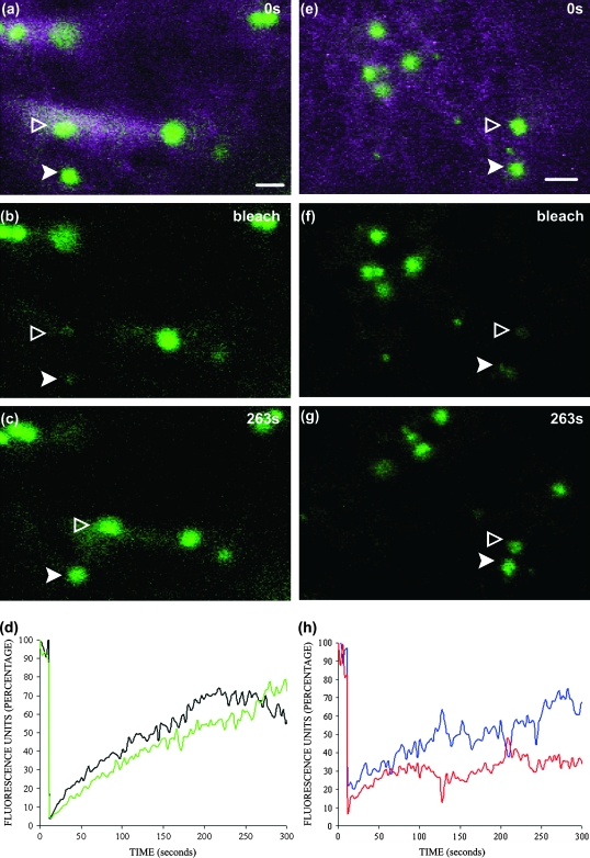
Although organelle movement in higher plants is predominantly actin-based, potential roles for the 17 predicted Arabidopsis myosins in motility are only just emerging. It is shown here that two Arabidopsis myosins from class XI, XIE, and XIK, are involved in Golgi, peroxisome, and mitochondrial movement. Expression of dominant negative forms of the myosin lacking the actin binding domain at the amino terminus perturb organelle motility, but do not completely inhibit movement. Latrunculin B, an actin destabilizing drug, inhibits organelle movement to a greater extent compared to the effects of AtXIE-T/XIK-T expression. Amino terminal YFP fusions to XIE-T and XIK-T are dispersed throughout the cytosol and do not completely decorate the organelles whose motility they affect. XIE-T and XIK-T do not affect the global actin architecture, but their movement and location is actin-dependent. The potential role of these truncated myosins as genetically encoded inhibitors of organelle movement is discussed.


Figures

Fig. 1.
Fig. 1.
Expression of eYFP-XIE-T or eYFP-XIK-T in tobacco leaf epidermal cells perturbs Golgi and peroxisome movement. A schematic to scale representation of AtXIE-T (a) and AtXIK-T (b) is shown and the tail domain is highlighted. Where the predicted myosin motor actin binding domain (green), IQ motif region (blue), coiled coil domains (yellow), and dilute region (red) are depicted. Movies of Golgi bodies (c, e, g) or peroxisomes (d, f, h) were generated and analysed using ‘Volocity’ tracking software. Tracks (lines, where each connecting point represents one time point in the movie sequence) from representative movies of each organelle (outlined objects) were generated and are shown in each panel. Cells co-expressing eYFP-XIE-T (e, f) or eYFP-XIK-T (g, h) and control cells only expressing organelle markers (c, d) are presented. The myosin tail fusion location is not shown. Expression of eYFP-XIK-T or eYFP-XIE-T severely perturbs Golgi and peroxisome movement over the time-course of the movie. Note, some organelles still display an obvious directional movement, albeit reduced, in the presence of eYFP-XIK-T or eYFP-XIE-T tail domain fusions (arrows). Scale bar 2 μm.
Fig. 2.
Fig. 2.
Statistical analysis of the effects of eYFP-XIE-T and eYFP-XIK-T on Golgi and peroxisome movement. Cumulative distribution function (CDF) plots of tracked peroxisomes (CFP-SKL, a, b, c) and Golgi bodies (ST-CFP, d, e, f) from at least 10 independent movies under various experimental conditions were generated. Organelle track velocity (a, d), displacement rates (shortest distance between the beginning and end of a track over time) (b, e), and meandering index (displacement rate divided by the track velocity) (c, f) are shown. Tracks from untreated control samples only expressing markers for Golgi bodies (n=543, dotted line) or peroxisomes (n=108, dotted line) are shown. Golgi body tracks from samples treated with Latrunculin B (n=106, dashed line) or co-expressing eYFP-XIE-T (n=243, black line) or eYFP-XIK-T (n=125, grey line) are slower and have lower rates than control samples, with Latrunculin B treatment having the lowest rates. Similar results are also apparent for peroxisome movement [peroxisome control (n=108), peroxisome sample treated with Latrunculin B (n=71), peroxisome sample co-expressing eYFP-XIE-T (n=52), or eYFP-XIK-T (n = 79)].
Fig. 3.
Fig. 3.
Expression of untagged XIE-T or XIK-T in tobacco epidermal cells perturbs Golgi body movement. Images taken from movies of cells expressing the Golgi body marker ST-CFP (a–c) or with untagged XIE-T (e–g) or untagged XIK-T (i–k) are shown. Note, global differences in Golgi positioning (black spots) over time in control cells (a–c) compared with Golgi location in cells expressing untagged XIE-T or XIK-T. Images generated during this time frame were subjected to ‘Volocity’ track analysis. The tracks generated for each organelle under control (d), XIE-T (h) or XIK-T (l) expression are shown, clearly indicating Golgi movement in control versus myosin tail expression are different. Time is shown in seconds on each panel; scale bar 5 μm.
Fig. 4.
Fig. 4.
Co-expression of eYFP-XIE-T or eYFP-XIK-T with various fluorescent organelle markers in tobacco epidermal cells. eYFP-XIE-T (magenta a, e, i, m) was co-expressed with fluorescent markers for Golgi bodies (ST-CFP, b), peroxisomes (CFP-SKL, f), mitochondria (β ATPase signal peptide-GFP, j), and the ER (GFP-HDEL, n). The merged images indicate that eYFP-XIE-T is closely associated, but does not solely decorate the periphery of Golgi bodies (c), peroxisomes (g), mitochondria (k), or the ER (o). Merged images of cells co-expressing eYFP-XIK-T (magenta) with fluorescent markers (green) for Golgi bodies (d), peroxisomes (h), mitochondria (l), and ER (p) also show a close association of eYFP-XIK-T with these organelles. Both eYFP-XIE-T and eYFP-XIK-T are present in large and small puncta. eYFP-XIE-T is in more numerous small puncta than eYFP-XIK-T, whereas eYFP-XIK-T is more diffuse throughout the cytosol than eYFP-XIE-T (compare c with d). Scale bar 5 μm.
Fig. 5.
Fig. 5.
Statistical analysis of eYFP-XIE-T and eYFP-XIK-T movement. CDF plots were generated of eYFP-XIE-T and eYFP-XIK-T large puncta tracks generated using ‘Volocity’ software. Track velocity (a), displacement rates (shortest distance between the beginning and end of a track over time, b), and meandering index (displacement rate divided by the track velocity, c) are shown. Tracks were generated from samples treated with Latrunculin B and from untreated cells. eYFP-XIK-T Latrunculin B treatment (n=39) black line, untreated cells (n=27) grey line, eYFP-XIE-T Latrunculin B treatment (n=27) black dashed line, and untreated cells (n=44) grey dotted line.
Fig. 6.
Fig. 6.
eYFP-XIE-T and eYFP-XIK-T association with the actin cytoskeleton. eYFP-XIE-T (a, b) and eYFP-XIK-T(c, d) were expressed in tobacco epidermal cells. Images were taken of cells treated with Latrunculin B (b, d) and control untreated cells (a, c). After treatment there are fewer small puncta, but the large puncta still remain. eYFP-XIE-T (magenta, e–g) and eYFP-XIK-T (magenta, h–j) were expressed with an actin marker, GFP-FABD2 (green, f, i). The global architecture of the actin appears unchanged by myosin tail expression, and the myosin tails puncta are frequently closely associated with the actin filaments. Scale bar 5 μm.
Fig. 7.
Fig. 7.
FRAP of ST-CFP in cells co-expressing eYFP-XIE-T or eYFP-XIK-T. ST-CFP within Golgi bodies (arrowheads) were bleached and recovery monitored over time in tobacco epidermal cells co-expressing either eYFP-XIE-T (a, b, c, d) or eYFP-XIK-T(e, f, g, h). Images shown were taken pre-bleach (a, e), immediately after bleaching (b, f) and 263 s after bleaching (c, g). Golgi bodies are still motile and so recovery of fluorescence is not at a steady rate due to movement of the organelle within the focal plane (graphs d and h). Curves are representative from at least five independent FRAP experiments.

References

    1. Allan VJ, Thompson HM, McNiven MA. Motoring around the Golgi. Nature Cell Biology. 2002;4:E236–E242. - PubMed
    1. Avisar D, Prokhnevsky AI, Makarova KS, Koonin EV, Dolja VV. Myosin XI-K is required for rapid trafficking of Golgi stacks, peroxisomes and mitochondria in leaf cells of Nicotiana benthamiana. Plant Physiology. 2008;146:1098–1108. - PMC - PubMed
    1. Batoko H, Zheng HQ, Hawes C, Moore I. A Rab1 GTPase is required for transport between the endoplasmic reticulum and Golgi apparatus and for normal Golgi movement in plants. The Plant Cell. 2000;12:2201–2218. - PMC - PubMed
    1. Boevink P, Oparka K, Santa-Cruz S, Martin B, Betteridge A, Hawes C. Stacks on tracks:the plant Golgi apparatus traffics on an actin/ER network. The Plant Journal. 1998;15:441–447. - PubMed
    1. Brandizzi F, Snapp EL, Roberts AG, Lippincott-Schwartz J, Hawes C. Membrane protein transport between the endoplasmic reticulum and the Golgi in tobacco leaves is energy-dependent but cytoskeleton-independent: evidence from selective photobleaching. The Plant Cell. 2002;14:1293–1309. - PMC - PubMed

Publication types

MeSH terms