. 2008 Sep;10(9):1866-78.
doi: 10.1111/j.1462-5822.2008.01177.x. Epub 2008 Jun 28.

ESX-1-dependent cytolysis in lysosome secretion and inflammasome activation during mycobacterial infection

Affiliations

ESX-1-dependent cytolysis in lysosome secretion and inflammasome activation during mycobacterial infection

Ingrid C Koo et al. Cell Microbiol. 2008 Sep.

Abstract

Exocytosis of lysosomes from macrophages has been described as a response to microbial cytotoxins and haemolysins, as well as for releasing pro-inflammatory cytokines interleukin (IL)-1beta and IL-18 during inflammasome activation. The mycobacterial ESX-1 secretion system, encoded in part by the Region of Difference-1, is a virulence factor necessary for phagosome escape and host cell lysis by a contact-dependent haemolysin in Mycobacterium marinum. Here we show that ESX-1 from M. marinum and M. tuberculosis is required for Ca(2+)-dependent induction of lysosome secretion from macrophages. Mycobacteria-induced lysosome secretion was concurrent to release of IL-1beta and IL-18, dependent on phagocytosis of bacteria containing ESX-1. Synthesis but not release of IL-1beta and IL-18 occurred in response to dead bacilli and bacteria lacking ESX-1, indicating that only cytokine release was regulated by ESX-1. Release of these cytokines and exocytosis of lysosomes were independent of intracellular mycobacterial growth, yet correlated with mycobacteria-encoded haemolytic activity, demonstrating a parallel pathway for the two responses. We further identified inflammasome components caspase-1, ASC and NALP3, but not Ipaf, required for release of IL-1beta and IL-18. Collectively, these results reveal a role for ESX-1 in triggering secretion of lysosomes, as well as release of IL-1beta and IL-18 during mycobacteria infection.


Figures

Figure 1
Figure 1
Mm triggers exocytosis of lysosomes at sites of invasion. (A) Confocal time-lapse microscopy of RFP-LAMP1 and MmGFP. Cells were visualized 5 minutes after inoculum was centrifuged on to monolayer. Minutes after the beginning of recording are indicated. Last frame shows outline of cell at end of recording. Arrows indicate new invading MmGFP at periphery of cell. (B) Staining of externalized LAMP1 after addition of MmGFP. Cells were fixed 5 minutes after addition of inoculum, and cells were immunostained for lumenal LAMP1 in the absence of cell permeabilization. Arrows indicate invading MmGFP in close proximity to exocytosed lysosomes.
Figure 2
Figure 2
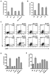
Mm-induced lysosome secretion depends on ESX-1 encoded hemolytic activity. (A) Secretion of lysosomes in response Mm strains (MOI = 50): no bacteria (NB), wild-type (WT), ESX-1 mutant (ΔRD1), heat-killed Mm (HK-Mm), or the complemented ESX-1 mutant (ΔRD1::RD1-2F9). (B) Secretion of lysosomes in response to Mtb strains (MOI = 50): no bacteria (NB), wild-type (Mtb), Rv3849::tn (11D6), or heat-killed Mtb (HK). (C) Flow cytometric analysis of LAMP1 externalization 10 minutes and 2 hours after infection by MmGFP, ΔRD1-GFP, and HK-GFP. Histogram shows GFP-positive cells stained for LAMP1 externalization. (D) Contact-dependent sheep erythrocyte hemolysis after incubation with Mm mutants. (E) Secretion of lysosomes in response to Mm mutants. Secreted β-hexosaminidase was measured from supernatants collected 2 hours after infection. All experiments were performed in triplicate.
Figure 3
Figure 3
Mm-induced lysosome secretion is regulated by intracellular Ca2+ and is necessary for host cell survival. (A) Effect of depletion of extracellular Ca2+ (no [Ca2+]e) or intracellular Ca2+ (BAPTA/AM) on secretion of β-hexosaminidase. (B) Measurement of cell death by induced by Mm in the absence of Ca2+. Cell death was measured by the CellTiter Blue assay (Promega). Asterisks (*) indicate levels beyond detection. Assays were performed on samples collected 2 hours after infection. All experiments were performed in triplicate.
Figure 4
Figure 4
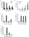
ESX-1 is necessary for secretion, but not synthesis of IL-1β and IL-18. (A) Release of IL-1β from BMDM in response to Mm strains: wild-type (WT), ESX-1 mutant (ΔRD1), and heat-killed Mm (HK). MOIs are indicated as bacteria:macrophage ratio. (B) IL-1β in lysates of infected BMDM. Asterisks (*) indicate levels below detection. (C) Release of IL-18 in response to Mm infection. (D) Secretion of TNF-α in response to Mm infection. (E) Release of IL-1β from BMDM in response to Mtb strains: wild-type (Erdman), Rv3849::tn, and heat-killed Mtb (HK). All assays were performed in triplicate, using supernatants and lysates collected 24 hours after infection.
Figure 5
Figure 5
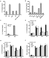
ESX-1 is necessary for triggering the release of IL-1β and IL-18 from primed BMDM. BMDM were primed with LPS, wild-type (WT), ESX-1 mutant (ΔRD1), or heat-killed (HK) Mm overnight. Cytokine release was measured 2 hrs after infection (panels A,C) or reinfection (panels B, D-F) using indicated Mm strains. (A) Release of IL-1β from LPS-primed BMDM in response to infection by Mm. (B) Release of IL-1β from LPS-primed BMDM in response to Mm mutants (C) Release of IL-1β from LPS-primed BMDM in the absence of phagocytosis. Cytochalasin D was used to treat cells prior to infection by Mm. (D) Stimulation of IL-1β release from Mm-primed BMDM in response to reinfection by Mm. (E) Release of IL-18 from Mm-primed BMDM in response to reinfection by Mm. (F) Secretion of TNF-α from Mm-primed BMDM in response to reinfection by Mm. All assays were performed in triplicate.
Figure 6
Figure 6
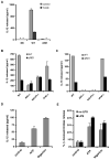
Mm activates the NALP3/ASC inflammasome. (A) Effect of caspase-1 inhibitor YVAD (50 μM) on secretion of IL-1β from BMDM. (B) Secretion of IL-1β from macrophages of ASC−/− and NALP3 knockout (NALP3−/−) mice. (C) Secretion of IL-18 from macrophages of ASC−/− and NALP3 knockout (NALP3−/−) mice. (D) Secretion of IL-1β from LPS-primed BMDM in response to ATP and nigericin. (E) Secretion of β-hexosaminidase from untreated or LPS-primed BMDM in response to ATP and nigericin. Assays were conducted using samples collected 2 hours after infection. Asterisks (*) indicate levels below detection.

References

    1. Andrei C, Dazzi C, Lotti L, Torrisi MR, Chimini G, Rubartelli A. The secretory route of the leaderless protein interleukin 1beta involves exocytosis of endolysosome-related vesicles. Mol Biol Cell. 1999;10:1463–1475. - PMC - PubMed
    1. Andrei C, Margiocco P, Poggi A, Lotti LV, Torrisi MR, Rubartelli A. Phospholipases C and A2 control lysosome-mediated IL-1 beta secretion: Implications for inflammatory processes. Proc Natl Acad Sci U S A. 2004;101:9745–9750. - PMC - PubMed
    1. Cox JS, Chen B, McNeil M, Jacobs WR., Jr Complex lipid determines tissue-specific replication of Mycobacterium tuberculosis in mice. Nature. 1999;402:79–83. - PubMed
    1. Czibener C, Sherer NM, Becker SM, Pypaert M, Hui E, Chapman ER, et al. Ca2+ and synaptotagmin VII-dependent delivery of lysosomal membrane to nascent phagosomes. J Cell Biol. 2006;174:997–1007. - PMC - PubMed
    1. de Jonge MI, Pehau-Arnaudet G, Fretz MM, Romain F, Bottai D, Brodin P, et al. ESAT-6 from Mycobacterium tuberculosis dissociates from its putative chaperone CFP-10 under acidic conditions and exhibits membrane-lysing activity. J Bacteriol 2007 - PMC - PubMed

Publication types

MeSH terms