. 2008 Sep 11;27(40):5373-84.
doi: 10.1038/onc.2008.155. Epub 2008 May 26.

Identification of VEGF-regulated genes associated with increased lung metastatic potential: functional involvement of tenascin-C in tumor growth and lung metastasis

Affiliations

Identification of VEGF-regulated genes associated with increased lung metastatic potential: functional involvement of tenascin-C in tumor growth and lung metastasis

A Calvo et al. Oncogene. .

Abstract

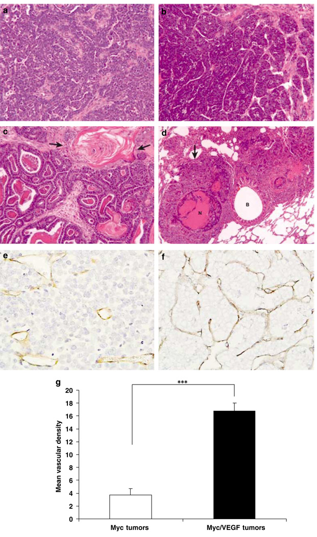
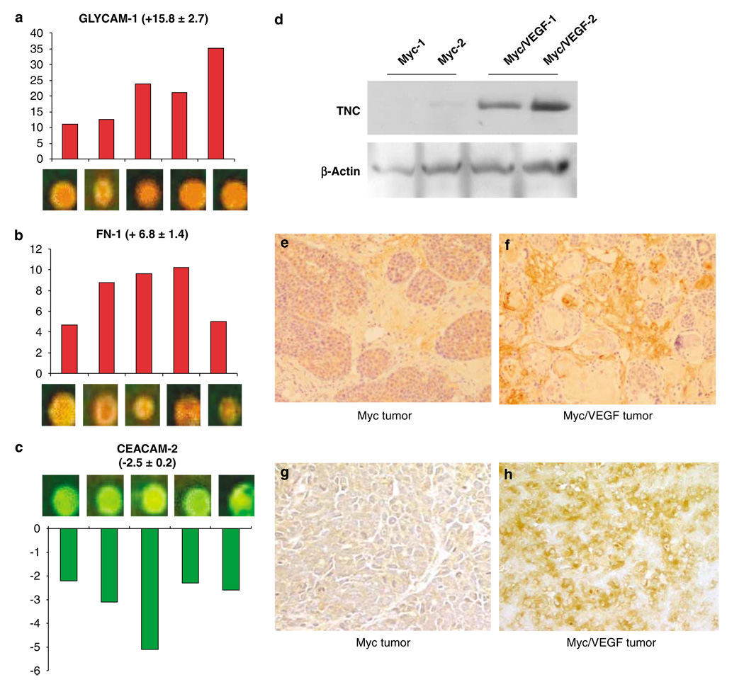
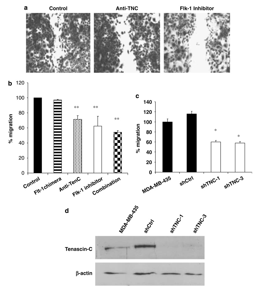
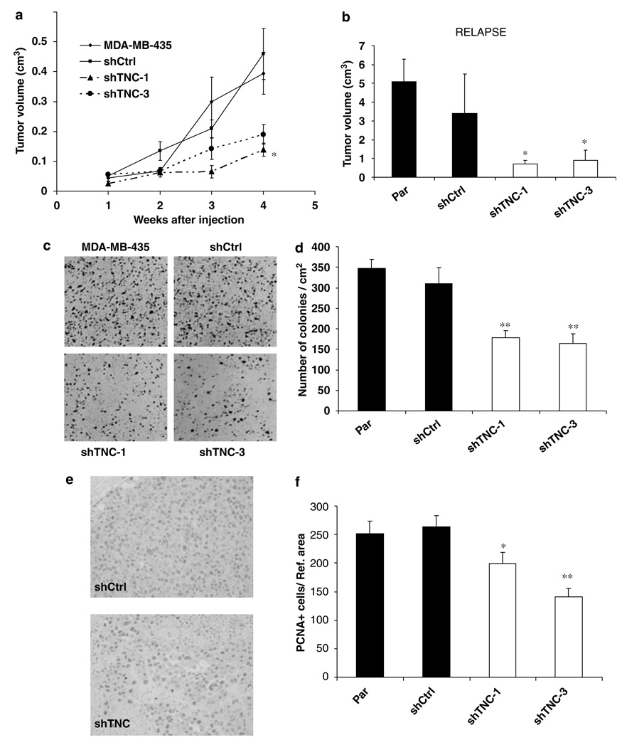
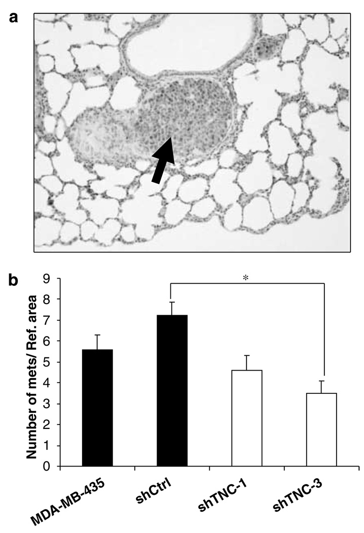
Metastasis is the primary cause of death in patients with breast cancer. Overexpression of c-myc in humans correlates with metastases, but transgenic mice only show low rates of micrometastases. We have generated transgenic mice that overexpress both c-myc and vascular endothelial growth factor (VEGF) (Myc/VEGF) in the mammary gland, which develop high rates of pulmonary macrometastases. Gene expression profiling revealed a set of deregulated genes in Myc/VEGF tumors compared to Myc tumors associated with the increased metastatic phenotype. Cross-comparisons between this set of genes with a human breast cancer lung metastasis gene signature identified five common targets: tenascin-C(TNC), matrix metalloprotease-2, collagen-6-A1, mannosidase-alpha-1A and HLA-DPA1. Signaling blockade or knockdown of TNC in MDA-MB-435 cells resulted in a significant impairment of cell migration and anchorage-independent cell proliferation. Mice injected with clonal MDA-MB-435 cells with reduced expression of TNC demonstrated a significant decrease (P<0.05) in (1) primary tumor growth; (2) tumor relapse after surgical removal of the primary tumor and (3) incidence of lung metastasis. Our results demonstrate that VEGF induces complex alterations in tissue architecture and gene expression. The TNC signaling pathway plays an important role in mammary tumor growth and metastases, suggesting that TNC may be a relevant target for therapy against metastatic breast cancer.


Figures

Figure 1
Figure 1
Morphology of primary Myc and Myc/VEGF mammary tumors. (a) Adenocarcinoma of a Myc transgenic mouse. (b) Adenocarcinoma of a Myc/VEGF mouse. (c) Adenosquamous (metaplastic) carcinoma of a Myc/VEGF mouse. Arrows indicate stratified squamous epithelium with keratinization. (d) Large pulmonary metastasis (arrow) with central necrosis (N). Metastasis surrounds a bronchus (B). (e) Immunostaining for CD-31 in a Myc tumor. (f) Myc/VEGF tumor showing higher vascularization (anti-CD-31 staining) than Myc tumors. (g) Comparison of the mean vascular density between Myc and Myc/VEGF tumors. ***P < 0.0004.
Figure 2
Figure 2
Changes in gene expression between Myc and Myc/VEGF tumors. (a) Microarray spots for GLYCAM-1 after competitive hybridization of Myc/VEGF tumors with Myc tumors. Bars represent fold-change increase in Myc/VEGF tumors compared to Myc tumors. (b) Microarray spots and fold-change increase for fibronectin-1 (FN-1) in Myc/VEGF tumors, as compared to Myc cancers. (c) CEACAM-2 is downregulated in bitransgenic tumors in comparison with Myc malignant tissues. (d) Validation of the upregulation of TCN by western blot. (e) Myc tumor stained for TNC. As seen by immunohistochemistry, TNC was mainly found in the extracellular matrix (ECM) and, in a lesser extent, in tumor cells. (f) Myc/VEGF tumor stained for TNC. Immunostaining for TNC is stronger in Myc/VEGF than in Myc tumors. (g) Myc tumor stained for laminin-5. Staining is observed in tumor cells and in the ECM. (h) Myc/VEGF tumor stained for laminin-5. A stronger staining was found for these types of tumors.
Figure 3
Figure 3
Migration assays. (a) Migration of M630 cells is impaired by TCN and Flk-1 signaling blockade. (b) Quantification of migration inhibition in M630 cells. Treatment with an Flt-1 antagonist did not change cell migration. Treatment with an Flk-1 inhibitor results in a significant reduction of migration (P < 0.01). Treatment with an anti-TNC blocking antibody also causes significant inhibition of cell migration (P < 0.01). The combination treatment using anti-TNC plus Flk-1 inhibitor produces the strongest inhibitory effect (P < 0.01). (c) Migration assays in MDA-MB-435 cells and cell clones shCtrl, shTNC-1 and shTNC-3. Downregulation of TNC in clones shTNC-1 and shTNC-3 reduces significantly (P < 0.05) cell migration when compared to MDA-MB-435 cells and the control clone shCtrl. (d) Western blot analysis to determine TNC protein expression in MDA-MB-435, shCtrl, shTNC-1 and shTNC-3 cells. *P < 0.05; **P < 0.01.
Figure 4
Figure 4
Decrease in primary tumor growth and cell proliferation in MDA-MB-435 cells with a downregulated expression of TNC. (a) Primary tumor volume is significantly lower (P < 0.05) in shTNC clones than in controls. (b) Relapsed tumor volume is significantly lower (P < 0.05) in cell clones lacking TCN. (c) Clonogenic assay in anchorage-independent conditions shows a lower number of colonies in shTNC clones, compared to controls. (d) Quantification of the number of colonies demonstrates that reduction in TNC levels results in lower clonogenic potential (P < 0.01) than that found for controls. (e) Immunohistochemistry for PCNA shows a decrease in PCNA-positive cells in shTNC tumors, compared to controls. (f) Quantification of PCNA-positive cells in primary tumors demonstrates that MDA-MB-435 and shCtrl tumors have similar rate of proliferating cells, which is higher than that found for shTCN-1 and shTNC-3 clones. *P < 0.05; **P < 0.01.
Figure 5
Figure 5
Downregulation of TNC in MDA-MB-435 cells reduces the number of lung metastatic foci. (a) Metastatic nodule (arrow) in the lung of a mouse injected with shCtrl cells. (b) Quantification of the number of lung metastatic foci. The average number of metastatic nudules was lower in animals injected with shTNC-3 cell clones than in mice injected with shCtrl cells (P < 0.05). No statistically significant differences were found when comparing data from shTNC-1 and shTNC-3 cells with parental cells or when comparing data from shTNC-1 with the shCtrl group (*P = 0.05 for the latter comparison).

References

    1. Adams J, Carder PJ, Downey S, Forbes MA, MacLennan K, Aligar V, et al. Vascular endothelial growth factor (VEGF) in breast cancer: comparison of plasma, serum, and tissue VEGF and microvessel density and effects of tamoxifen. Cancer Res. 2000;60:2898–2905. - PubMed
    1. Amundadottir LT, Johnson MD, Merlino G, Smith GH, Dickson RB. Synergistic interaction of transforming growth factor alpha and c-myc in mouse mammary and salivary gland tumorigenesis. Cell Growth Differ. 1995;6:737–748. - PubMed
    1. Calvo A, Xiao N, Kang J, Best CJ, Leiva I, Emmert-Buck MR, et al. Alterations in gene expression profiles during prostate cancer progression: functional correlations to tumorigenicity and down-regulation of selenoprotein-P in mouse and human tumors. Cancer Res. 2002;62:5325–5335. - PubMed
    1. Chiquet-Ehrismann R, Chiquet M. Tenascins: regulation and putative functions during pathological stress. J Pathol. 2003;200:488–499. - PubMed
    1. De Paola F, Granato AM, Scarpi E, Monti F, Medri L, Bianchi S, et al. Vascular endothelial growth factor and prognosis in patients with node-negative breast cancer. Int J Cancer. 2002;98:228–233. - PubMed

Publication types

MeSH terms