. 2008 Aug 28;27(37):5057-68.
doi: 10.1038/onc.2008.143. Epub 2008 May 26.

Mitochondrial redox signaling by p66Shc is involved in regulating androgenic growth stimulation of human prostate cancer cells

Affiliations

Mitochondrial redox signaling by p66Shc is involved in regulating androgenic growth stimulation of human prostate cancer cells

S Veeramani et al. Oncogene. .

Abstract

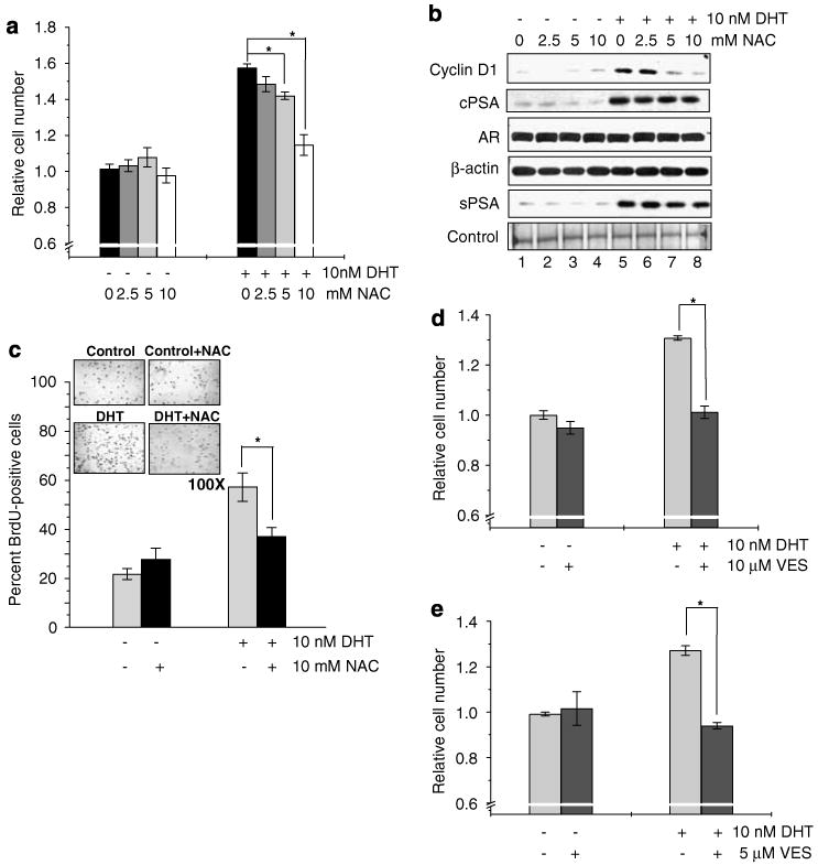
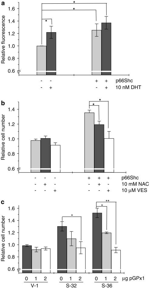
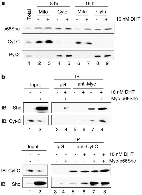
p66Shc is shown to negatively regulate the life span in mice through reactive oxygen species (ROS) production. Recent reports, however, revealed that p66Shc protein level is significantly elevated in several human cancer tissues and growth-stimulated carcinoma cells, suggesting a mitogenic and carcinogenic role for p66Shc. In this communication, we demonstrate for the first time that p66Shc mediates androgenic growth signals in androgen-sensitive human prostate cancer cells through mitochondrial ROS production. Growth stimulation of prostate cancer cells with 5alpha-dihydrotestosterone (DHT) is accompanied by increased p66Shc level and ROS production, which is abolished by antioxidant treatments. However, antioxidant treatments do not affect the transcriptional activity of androgen receptor (AR) as observed by its inability to block DHT-induced prostate-specific antigen expression, an AR-dependent correlate of prostate cancer progression. Elevated expression of p66Shc by cDNA transfection increases the basal cell proliferation and, thus, reduces additional DHT-induced cell proliferation. Furthermore, DHT increases the translocation of p66Shc into mitochondria and its interaction with cytochrome c. Conversely, both redox-negative p66Shc mutant (W134F), which is deficient in cytochrome c interaction, and p66Shc small interfering RNA decrease DHT-induced cell proliferation. These results collectively reveal a novel role for p66Shc-ROS pathway in androgen-induced prostate cancer cell proliferation and, thus, may play a role in early prostate carcinogenesis.


Figures

Figure 1
Figure 1
Requirement of reactive oxygen species (ROS) production in dihydrotestosterone (DHT)-induced prostate cancer cell proliferation. (a) LNCaP cells were plated at a density of 1 × 10⁴ cells per cm2 in regular medium. Seventy-two hours later, cells were steroid starved using steroid-reduced medium for 48 h. Cells were then treated with 10 nM DHT in the presence or absence of different doses of N-acetyl cysteine (NAC) (0–10mM) for 48 h. Cell growth was determined by cell counting (*P<0.01; r = −0.988, P<0.05). (b) Immunoblot analysis of Cyclin D1, androgen receptor (AR), prostate-specific antigen (PSA) levels in cell lysates (cPSA) and secreted PSA in condition medium (sPSA) was performed in DHT-treated LNCaP cells in the presence or absence of NAC (0–10 mM). (c) DHT-treated LNCaP cells in the presence or absence of 10mM NAC were immunostained for bromodeoxyuridine (BrdU) incorporation, which showed inhibition of DHT-induced BrdU incorporation by antioxidant (*P<0.01). (d) LNCaP cell growth induced by 10 nM DHT was abolished by 10 μM vitamin E succinate (VES), another antioxidant (*P<0.01). (e) MDA PCa2b cells were plated at a density of 4 × 10⁴ cells per cm2 and were allowed to attach for 3 days. Cells were then starved with steroid-reduced medium, as mentioned in the Methods section, for 48 h. Cells were treated with DHT in the presence or absence of VES (10 μM) and cell growth was measured by cell counting after 72 h (*P<0.01).
Figure 2
Figure 2
Stimulation of proliferation of prostate cancer cells by increased intracellular reactive oxygen species (ROS) levels. (a) LNCaP cells plated at a density of 1 × 10⁴ cells per cm2 were steroid starved for 48 h and, later, were treated with different doses of H2O2 (0–20 μm) or 10 nM dihydrotestosterone (DHT) for 48 h. Cell growth was determined by cell counting (*P<0.05; **P<0.01). (b) Immunoblot analysis was performed for Cyclin D1, androgen receptor (AR), prostate-specific antigen (PSA) levels in cell lysates (cPSA) and secreted PSA levels in conditioned medium (sPSA) in LNCaP cells treated with different doses of H2O2 (0–20 μM) or 10 nM DHT. (c) LNCaP cells were plated at a density of 1 × 10⁴ cells per cm2 for 48 h and transfected with superoxide dismutase (SOD) cDNA. Control cells were transfected with empty vector. Cells were steroid starved for 48 h and then were treated with ethanol or 10 nM DHT. Cells were counted 48 h post-treatment (*P<0.05). (d) Immunoblot analysis was performed in SOD-overexpressed LNCaP cells for Cyclin D1 and PSA levels in cell lysates.
Figure 3
Figure 3
Elevation of p66Shc prior to reactive oxygen species (ROS) production and cell growth induction in dihydrotestosterone (DHT)-treated LNCaP cells. LNCaP cells were plated and steroid starved for 48 h. Cells were then treated with 10 nM DHT and control cells were treated with equal volume of ethanol. Cells were harvested at specific time periods and were analysed for (a) Shc isoforms, Cyclin D1 and secreted prostate-specific antigen (sPSA) protein levels by immunoblotting, (b) ROS production using DCF-DA analysis (*P<0.05), (c) cell cycle analysis (*P<0.01) by flow cytometry and (d) cell growth analysis (*P<0.01) by cell counting.
Figure 4
Figure 4
Stimulation of cell proliferation by overexpression of p66Shc in prostate cancer cells. (a) LNCaP cells were plated at a cell density of 1 × 10⁴ cells per cm2 for 48 h and then transfected with p66Shc wild-type cDNA. Control cells were transfected with empty vector. Transfected cells were starved for 48 h and then treated with ethanol or 10 nM dihydrotestosterone (DHT) for 48 h. Cell growth was analysed by cell counting (*P<0.01). (b) Immunoblot analysis was performed for Cyclin D1 protein levels in p66Shc-overexpressed LNCaP cells (lanes 3 and 4) and vector-alone transfected control cells (lanes 1 and 2), in the presence or absence of DHT. (c) LNCaP cells stably transfected with p66Shc cDNA (S-32 and S-36) and with vector-alone cells (V-1) were plated at a density of 8 × 10³ cells per cm2 in regular medium for 3 days. Cells were steroid starved for 48 h and then replenished with steroid-reduced medium. One set of the cells were trypsinized and counted 3 days later. The other set of cells were fed with fresh steroid-reduced medium and trypsinized at day 6 (*P<0.05). (d) Immunoblot analysis was performed for Cyclin D1 and p66Shc levels in p66Shc stable subclone cells that were harvested at day 3 of cell growth experiment. (e) MDA PCa2b cells were plated at a cell density of 4 × 10⁴ cells per cm2 for 48 h and were transfected with p66Shc wild-type cDNA or empty vector. Cells were steroid starved for 48 h and then treated with 10 nM DHT. Cell growth was analysed 72 h after DHT treatment by cell counting (*P<0.05).
Figure 5
Figure 5
Generation of reactive oxygen species (ROS) in p66Shc overexpressed prostate cancer cells. (a) LNCaP cells were plated at a density of 1 × 10⁴ cells per cm2 and co-transfected with p66Shc cDNA and enhanced cyan fluorescent protein (ECFP) cDNA. Control cells were transfected with empty vector and ECFP cDNA. After 48 h of steroid starvation, cells were treated with 10 nM dihydrotestosterone (DHT) or ethanol. DCF-DA assay was performed with 20 μM of DCF-DA in dimethyl sulfoxide for 15 min. Green DCF-DA fluorescence was measured in ECFP-positive cells by flow cytometry (*P<0.05). (b) LNCaP cells were transiently transfected as above and were steroid starved for 48 h. Cells were then treated with 10 mM N-acetyl cysteine (NAC) or 10 μM vitamin E succinate (VES) for additional 48 h and cell growth was measured (*P<0.05). (c) LNCaP cells stably transfected with p66Shc cDNA (S-32 and S-36) and with vector-alone cells (V-1) were plated at a density of 1 × 10⁴ cells per cm2 and were transfected with different amounts of cDNA encoding glutathione peroxidase 1 (pGPx1). Cells were then steroid starved for 48 h and then replenished with steroid-reduced medium. Forty-eight hours later cells were trypsinized and counted (*P<0.05, **P<0.01).
Figure 6
Figure 6
Translocation of p66Shc into mitochondria and its interaction with cytochrome c (Cyt c) in dihydrotestosterone (DHT)-treated LNCaP cells. (a) LNCaP cells were treated with 10 nM DHT and were subfractionated after 8 and 16 h after treatment as described (Giorgio et al., 2005). Total cell lysate (total) and the subfractionated mitochondrial (Mito) and cytosolic (Cyto) fractions were analysed by immunoblotting for p66Shc compartmentalization. Cyt c was used as a mitochondria-specific marker and Pyk2, a cytoplasmic tyrosine kinase, was used a cytosol-specific marker. (b) LNCaP cells were transiently transfected with Myc-tagged p66Shc wild-type cDNA or empty vector and steroid starved for 48 h. Cells were the treated with 10 nM DHT for 16 h. Co-immunoprecipitation assay was performed with anti-Myc antibody (as mentioned in the Materials and methods section) to analyse the interaction between Cyt c and exogenous p66Shc. Interaction was compared between p66Shc-overexpressed cells, which express both endogenous and exogenous p66Shc, in the presence or absence of DHT (upper panel, lanes 7 vs 8). To analyse the endogenous protein interaction, anti-Cyt c antibody was used to pull-down Cyt c and the membrane was probed was anti-Shc antibody. Interaction was compared between empty vector-transfected cells with or without DHT, which express only endogenous p66Shc (lower panel, lanes 5 vs 6).
Figure 7
Figure 7
Requirement of reactive oxygen species (ROS) production by p66Shc in dihydrotestosterone (DHT)-induced LNCaP cell proliferation. (a) LNCaP cells were transfected with wild-type p66Shc cDNA (WT) or W134F mutant or empty vector alone (pcDNA). Cells were steroid starved for 48 h and treated with 10 nM DHT as described above. Cells were trypsinized and counted after 48 h of treatment (*P<0.05). (b) Immunoblot analysis was performed for Cyclin D1 level in LNCaP cells transiently overexpressing WT or W134F mutant in the presence or absence of DHT, as mentioned above. (c) MDA PCa2b cells were plated at 4 × 10⁴ cells per cm2 and transiently transfected WT and W134F mutant of p66Shc and treated with DHT, as mentioned earlier. Seventy-two hours after DHT treatment cells were trypsinized and counted (*P<0.05). (d) Small interfering RNA (siRNA) fragments specific to p66Shc cloned in pSUPER vector (pSUP-p66) was transiently transfected into LNCaP cells as per the conditions mentioned for cDNA transfection. Cells were treated with DHT for 48 h and cell growth was measured (*P<0.01). (e) Immunoblot analysis was performed for Cyclin D1 levels in LNCaP cells transiently transfected with p66Shc-specific siRNA (pSUP-p66).
Figure 8
Figure 8
A schematic representation of the p66Shc pathway in androgen action on prostate cancer cell proliferation. Dihydrotestosterone (DHT)-bound androgen receptor (AR) complex can activate two independent pathways—AR-dependent gene expression, such as prostate-specific antigen (PSA), and AR-dependent cell proliferation, involving the p66Shc–reactive oxygen species (ROS) pathway. Stimulation of androgenic proliferation includes an increase in p66Shc protein level after DHT treatment. Increased p66Shc translocates into mitochondria, where it interacts with cytochrome c (Cyt c), localized in the mitochondrial inner membrane, and generates ROS production. Increased ROS production promotes cell cycle progression, through Cyclin D1 and, which in turn, increases cell proliferation. In addition to the activation of p66Shc–ROS pathway, a part of the cell growth is also mediated by the genomic effects of the AR-dependent pathway (Dotted arrow).

References

    1. Bourdon JC. p53 and its isoforms in cancer. Br J Cancer. 2007;97:277–282. - PMC - PubMed
    1. Davol PA, Bagdasaryan R, Elfenbein GJ, Maizel AL, Frackelton AR., Jr Shc proteins are strong, independent prognostic markers for both node-negative and node-positive primary breast cancer. Cancer Res. 2003;63:6772–6783. - PubMed
    1. Giorgio M, Migliaccio E, Orsini F, Paolucci D, Moroni M, Contursi C, et al. Electron transfer between cytochrome c and p66Shc generates reactive oxygen species that trigger mitochondrial apoptosis. Cell. 2005;122:221–233. - PubMed
    1. Grossman SR, Lyle S, Resnick MB, Sabo E, Lis RT, Rosinha E, et al. p66 Shc Tumor Levels Show a Strong Prognostic Correlation with Disease Outcome in Stage IIA Colon Cancer. Clin Cancer Res. 2007;13:5798–5804. - PubMed
    1. Horoszewicz JS, Leong SS, Kawinski E, Karr JP, Rosenthal H, Chu TM, et al. LNCaP model of human prostatic carcinoma. Cancer Res. 1983;43:1809–1818. - PubMed

Publication types

MeSH terms