Review
. 2008 Jun;18(6):609-21.
doi: 10.1038/cr.2008.61.

Feedback regulation of cholesterol synthesis: sterol-accelerated ubiquitination and degradation of HMG CoA reductase

Affiliations
Review

Feedback regulation of cholesterol synthesis: sterol-accelerated ubiquitination and degradation of HMG CoA reductase

Russell A DeBose-Boyd. Cell Res. 2008 Jun.

Abstract

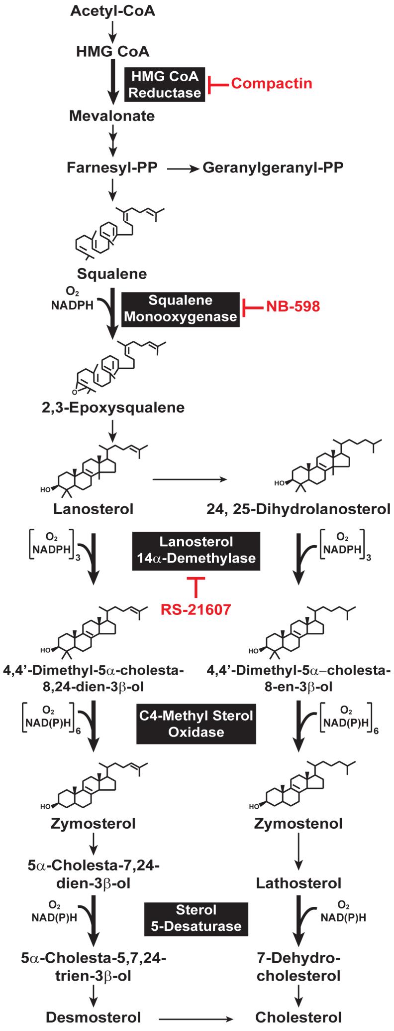
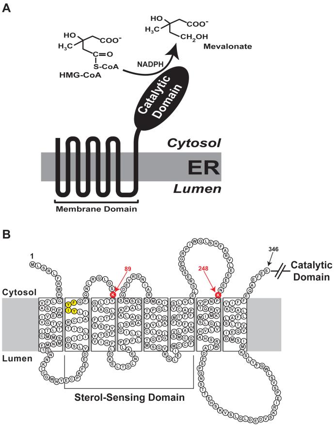
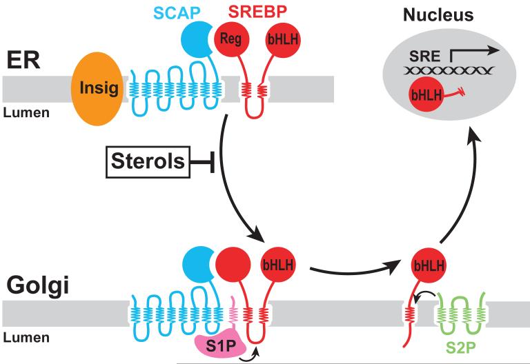
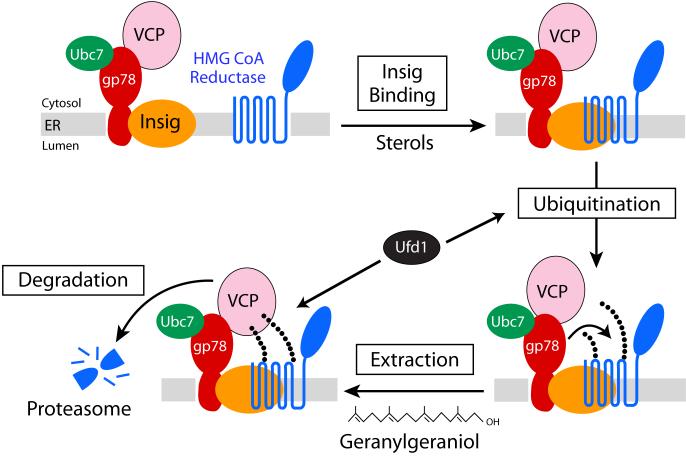
3-hydroxy-3-methylglutaryl coenzyme A (HMG CoA) reductase produces mevalonate, an important intermediate in the synthesis of cholesterol and essential nonsterol isoprenoids. The reductase is subject to an exorbitant amount of feedback control through multiple mechanisms that are mediated by sterol and nonsterol end-products of mevalonate metabolism. Here, I will discuss recent advances that shed light on one mechanism for control of reductase, which involves rapid degradation of the enzyme. Accumulation of certain sterols triggers binding of reductase to endoplasmic reticulum (ER) membrane proteins called Insig-1 and Insig-2. Reductase-Insig binding results in recruitment of a membrane-associated ubiquitin ligase called gp78, which initiates ubiquitination of reductase. This ubiquitination is an obligatory reaction for recognition and degradation of reductase from ER membranes by cytosolic 26S proteasomes. Thus, sterol-accelerated degradation of reductase represents an example of how a general cellular process (ER-associated degradation) is used to control an important metabolic pathway (cholesterol synthesis).


Figures

Figure 1
Figure 1
Schematic Representation of the Cholesterol Synthetic Pathway in Animal Cells. Reactions that require molecular oxygen are indicated, and specific inhibitors of various enzymes in the pathway are highlighted in red.
Figure 2
Figure 2
Domain Structure of Hamster HMG CoA Reductase. (A) HMG CoA reductase consists of two distinct domains: a hydrophobic N-terminal domain with eight membrane-spanning segments that anchor the protein to ER membranes; and a hydrophilic C-terminal domain that projects into the cytosol and exhibits all of the enzyme’s catalytic activity. (B) Amino acid sequence and topology of the membrane domain of hamster HMG CoA reductase. The lysine residues implicated as sites of Insig-dependent, sterol-regulated ubiquitination are highlighted in red and denoted by arrows. The YIYF sequence in the second membrane-spanning helix that mediates Insig binding is highlighted in yellow.
Figure 3
Figure 3
Model for Sterol-Regulated Scap-SREBP Pathway. SCAP is a sensor of sterols and an escort of SREBPs. In sterol-depleted cells, Scap facilitates export of SREBPs from the ER to the Golgi apparatus, where two proteases, Site-1 protease (S1P) and Site-2 protease (S2P), act to release the transcriptionally active, N-terminal bHLH-Zip domain of SREBPs from the membrane. The released bHLH-Zip domain migrates into the nucleus and binds to a sterol response element (SRE) in the enhancer/promoter region of target genes, activating their transcription. Accumulation of sterols in ER membranes trigger binding of Scap to one of two retention proteins called Insigs, which blocks incorporation of Scap-SREBP complexes into ER transport vesicles. As a result, SREBPs no longer translocate to the Golgi apparatus, the bHLH-Zip domain cannot be released from the membrane, and transcription of all target genes declines.
Figure 4
Figure 4
Mechanism for Oxygen Sensing in the Cholesterol Synthetic Pathway The link between synthesis of cholesterol and oxygen sensing in animal cells is provided by hypoxia induced accumulation of lanosterol and 24,25-dihydrolanosterol and HIF-1a-mediated induction of Insig-1 and Insig-2. Convergence of these responses leads to rapid degradation of HMG CoA reductase, thereby limiting synthesis of cholesterol.
Figure 5
Figure 5
Pathway for Sterol-Accelerated Degradation of HMG CoA Reductase Accumulation of 25-hydroxycholesterol, lanosterol, or 24,25-dihydrolanosterol in ER membranes triggers binding of the reductase to Insigs. A subset of Insigs is associated with the membrane-anchored ubiquitin ligase, gp78, which binds the E2 Ubc7 and VCP, an ATPase that plays a role in extraction of ubiquitinated proteins from ER membranes. Through the action of gp78 and Ubc7, reductase becomes ubiquitinated, which triggers its extraction from the membrane by VCP, and subsequent delivery to proteasomes for degradation. The post-ubiquitination step is postulated to be enhanced by geranylgeraniol through an undefined mechanism that may involve a geranylgeranylated protein, such as one of the Rab proteins.

References

    1. Goldstein JL, Brown MS. Regulation of the mevalonate pathway. Nature. 1990;343:425–430. - PubMed
    1. Brown MS, Goldstein JL. Multivalent feedback regulation of HMG CoA reductase, a control mechanism coordinating isoprenoid synthesis and cell growth. J. Lipid Res. 1980;21:505–517. - PubMed
    1. Endo A, Kuroda M, Tanzawa K. Competitive inhibition of 3-hydroxy-3-methylglutaryl coenzyme A reductase by ML-236A and ML-236B fungal metabolites, having hypocholesterolemic activity. FEBS Lett. 1976;72:323–326. - PubMed
    1. Nakanishi M, Goldstein JL, Brown MS. Multivalent control of 3-hydroxy-3-methylglutaryl coenzyme A reductase. Mevalonate-derived product inhibits translation of mRNA and accelerates degradation of enzyme. J. Biol. Chem. 1988;263:8929–8937. - PubMed
    1. Horton JD, Goldstein JL, Brown MS. SREBPs: activators of the complete program of cholesterol and fatty acid synthesis in the liver. J. Clin. Invest. 2002;109:1125–1131. - PMC - PubMed

Publication types