. 2007 Aug;3(3):255-68.
doi: 10.1017/S1740925X08000100.

Do glial cells control pain?

Affiliations

Do glial cells control pain?

Marc R Suter et al. Neuron Glia Biol. 2007 Aug.

Abstract

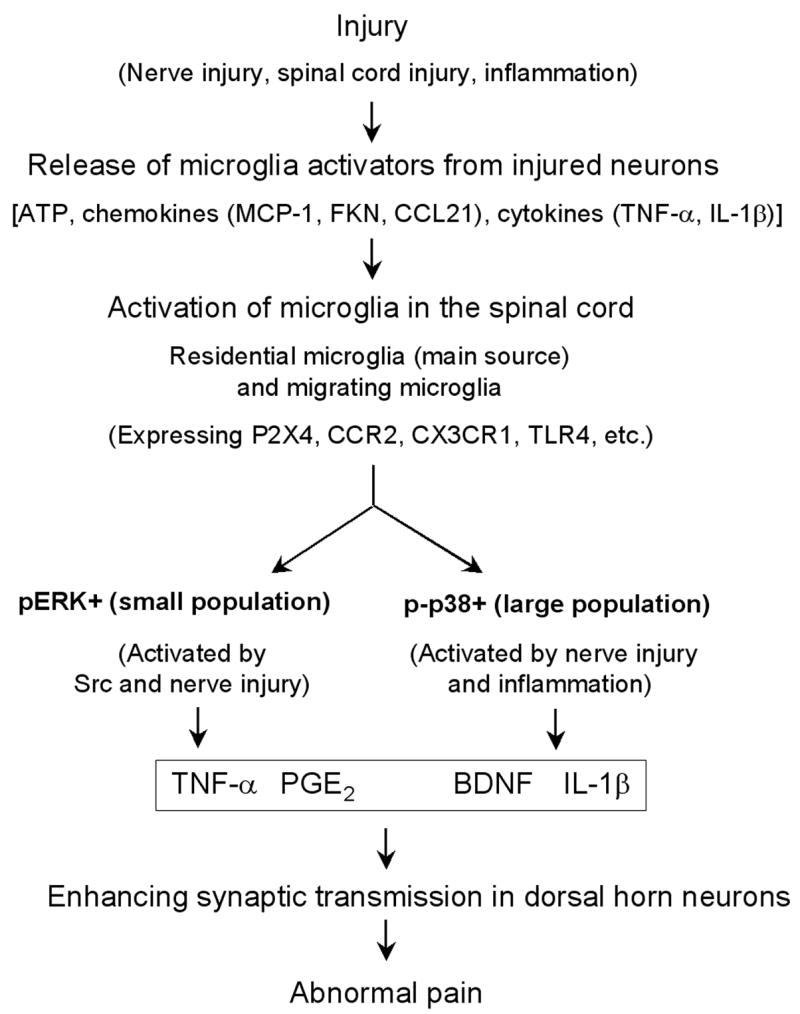
Management of chronic pain is a real challenge, and current treatments that focus on blocking neurotransmission in the pain pathway have resulted in limited success. Activation of glial cells has been widely implicated in neuroinflammation in the CNS, leading to neurodegeneration in conditions such as Alzheimer's disease and multiple sclerosis. The inflammatory mediators released by activated glial cells, such as tumor necrosis factor-a and interleukin-1b not only cause neurodegeneration in these disease conditions, but also cause abnormal pain by acting on spinal cord dorsal horn neurons in injury conditions. Pain can also be potentiated by growth factors such as brain-derived growth factor and basic fibroblast growth factor, which are produced by glia to protect neurons. Thus, glial cells can powerfully control pain when they are activated to produce various pain mediators. We review accumulating evidence that supports an important role for microglial cells in the spinal cord for pain control under injury conditions (e.g. nerve injury). We also discuss possible signaling mechanisms, in particular mitogen-activated protein kinase pathways that are crucial for glial-mediated control of pain.Investigating signaling mechanisms in microglia might lead to more effective management of devastating chronic pain.

Keywords: MAP kinase; chemokines; chronic pain; cytokines; intracellular signaling; microglia; nerve injury.


Figures

Figure 1
Figure 1. (a–d). Spared nerve injury (SNI) induces proliferation of microglial cells in the spinal cord
(a) Sham control (Cont) animals show almost no BrdU immunostaining. (b) Three days following SNI, there is a profound proliferation in the ipsilateral dorsal horn. Scale, 100 μm. (c) Double immunostaining of BrdU with the microglial marker Iba1 in the dorsal horn. Note that most BrdU positive cells also express Iba1 (indicated with arrows). White lines show the borders of the dorsal horn. Scale, 100 μm. (d) Number of BrdU-positive cells (per 30 μm-thick section) in the spinal cord dorsal horn. Note that proliferation starts rapidly after SNI.
Figure 2
Figure 2. (a–d). SNI induces CD11b (OX-42) upregulation in the spinal cord dorsal horn area that is terminated by injured nerve branches
(a, b) Isolectin B4 (IB4) staining for primary afferents shows a reduction of the staining in the medial ipsilateral dorsal horn (L4 level), an area terminated by the injured branches (tibial and peroneal) of the sciatic nerve, at 3 days after SNI. (c, d) Immunohistochemistry shows increased expression of OX-42 in the medial dorsal horn on the ipsilateral side 3 days after SNI. White lines indicate the borders of the dorsal horn. White stars indicate the spinal area terminated by the intact sural nerve. Scale, 100 μm. Modified from Wen et al., 2007.
Figure 3
Figure 3. (a–e). Activation of p38 and ERK in the spinal cord after SNI is required for neuropathic pain development
(a–d) Immunohistochemistry shows an increase in phosphorylation of p38 (p-p38, a, b) and ERK (pERK, c, d) in the medial dorsal horn on the ipsilateral side (L4) 3 days after SNI. White lines indicate the borders of the dorsal horn. Scales, 100 μm. (e) SNI-induced mechanical allodynia is prevented by the p38 inhibitor FR167653 or the MEK (ERK kinase) inhibitor U0126. FR167653 (30 μg/μl) or U0126 (1 μg/μl) was infused into intrathecal space via an osmotic pump (0.5 μl/h for 5 days) starting 2 days before SNI. Note that the basal mechanical sensitivity does not change after FR167653 or U0126 infusion. **, P < 0.01, t test, compared to corresponding vehicle controls (30% DMSO), n = 4. Modified from Wen et al., 2007.
Figure 4
Figure 4. (a–g). p38 and ERK are activated in different populations of microglia in the spinal cord following SNI
(a–f) Double immunofluorescence shows colocalization of p-p38 and OX-42 (a–c) and colocalization of pERK and OX-42 (d–f) in the medial superficial dorsal horn. (g) Double immunofluorescence shows that p-p38 and pERK are not activated in the same cells. c is an overlay of a and b, f is an overlay of d and e. Scales, 50 μm.
Figure 5
Figure 5. (a–c). Spinal nerve ligation (SNL) increases NF-κB expression in spinal microglia
(a, b) SNL increases the immunoreactivity of NF-κB (65kD unit) on day 3 in the medial superficial spinal cord, compared to non-injured control (Cont) spinal cord. Scale, 50 μm. (c) Double immunofluorescence shows colocalization of NF-κB and OX-42. Scale, 20 μm.
Figure 6
Figure 6. (a–c). Schematic representation of microglial control of pain
After various injury conditions (such as nerve injury, spinal cord injury, and inflammation), injured or affected neurons (e.g., DRG or spinal cord neurons) can release factors that are capable of activating microglia in the spinal cord. These factors include ATP, chemokines such as MCP-1, fractalkine (FKN), and CCL21, and proinflammatory cytokines such as TNF-α and IL-1β. These microglia activators can bind their receptors on microglia, leading to the activation of microglia. These activated spinal microglia are mainly residential microglia, but may also be migrating microglia from circulation. Activated microglia contain three subpopulations in the spinal cord: pERK + population, p-p38 + population, and unknown population. While ERK is activated by Src and nerve injury, p38 is activated by both nerve injury and inflammation. ERK activation releases TNF-α via activation of TNF-α converting enzyme, whereas p38 activation enhances IL-1β release. Activation of p38 and ERK can also regulate the synthesis of the inflammatory mediators via transcription factor (e.g., NF-κB). Microglia also produce the growth factor BDNF. Finally, these pain mediators collaborate to induce and maintain abnormal pain in injury conditions by enhancing synaptic strength and central sensitization in dorsal horn neurons.

References

    1. Abbadie C, Lindia JA, Cumiskey AM, Peterson LB, Mudgett JS, Bayne EK, DeMartino JA, MacIntyre DE, Forrest MJ. Impaired neuropathic pain responses in mice lacking the chemokine receptor CCR2. Proc Natl Acad Sci USA. 2003;100(13):7947–7952. - PMC - PubMed
    1. Aumeerally N, Allen G, Sawynok J. Glutamate-evoked release of adenosine and regulation of peripheral nociception. Neuroscience. 2004;127(1):1–11. - PubMed
    1. Bhat NR, Zhang P, Lee JC, Hogan EL. Extracellular signal-regulated kinase and p38 subgroups of mitogen-activated protein kinases regulate inducible nitric oxide synthase and tumor necrosis factor-alpha gene expression in endotoxin-stimulated primary glial cultures. J Neurosci. 1998;18(5):1633–1641. - PMC - PubMed
    1. Block ML, Zecca L, Hong JS. Microglia-mediated neurotoxicity: uncovering the molecular mechanisms. Nat Rev Neurosci. 2007;8(1):57–69. - PubMed
    1. Boyle DL, Jones TL, Hammaker D, Svensson CI, Rosengren S, Albani S, Sorkin L, Firestein GS. Regulation of peripheral inflammation by spinal p38 MAP kinase in rats. PLoS Med. 2006;3(9):e338. - PMC - PubMed

LinkOut - more resources