. 2008 Aug;23(8 Pt 2):e477-86.
doi: 10.1111/j.1440-1746.2008.05339.x. Epub 2008 May 26.

Insulin resistance in experimental alcohol-induced liver disease

Affiliations

Insulin resistance in experimental alcohol-induced liver disease

Suzanne M de la Monte et al. J Gastroenterol Hepatol. 2008 Aug.

Abstract

Background and aim: Chronic ethanol consumption impairs liver regeneration due, in part, to inhibition of insulin signaling. This study characterizes the mechanisms and consequences of ethanol-impaired insulin signaling in relation to oxidative injury and altered gene expression.

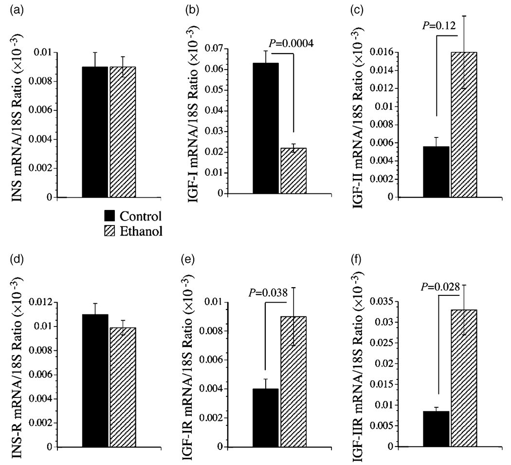
Methods: Long-Evans rats were fed for 8 weeks with isocaloric liquid diets containing 0% (control) or 37% ethanol (caloric content). Livers were used to examine histopathology, indices of oxidative stress, gene expression required for insulin and insulin-like growth factor (IGF) signaling, insulin-responsive gene expression, i.e. glyceraldehydes-3-phosphate dehydrogenase (GAPDH) and aspartyl-asparaginyl-beta-hydroxylase (AAH), and competitive equilibrium binding to the insulin, IGF-I, and IGF-II receptors.

Results: Chronic ethanol exposure caused liver injury with increased hepatocellular steatosis, inflammation, apoptosis, and increased immunoreactivity for activated caspase-3, 8-hydroxy-2'-deoxyguanosine, and 4-hydroxy-2,3-nonenol. These effects were associated with increased expression of IGF-I receptor, IGF-II, and IGF-II receptor, and expression of IGF-I, AAH, and GAPDH, which mediate energy metabolism and cell motility/remodeling, and reduced binding to the insulin receptor.

Conclusions: Chronic ethanol-induced liver injury causes insulin resistance with inhibition of insulin-responsive genes needed for metabolism, remodeling, and regeneration. In contrast, the IGF-I and IGF-II signaling mechanisms remain relatively preserved, suggesting that insulin-regulated hepatic functions may be selectively vulnerable to the toxic effects of ethanol.


Figures

Figure 1
Figure 1
Histopathology of chronic ethanol-induced liver injury. Adult Long-Evans rats were fed for 8 weeks with isocaloric liquid diets containing 0% (n = 12) (a,c,e) or 37% (n = 13) (b,d,f) ethanol by caloric content. Paraffin-embedded sections of liver were stained with hematoxylin–eosin (a,b). Note the disordered hepatic architecture with loss of chord organization and increased steatosis (inset) in the ethanol-exposed liver. Adjacent sections were immunostained to detect 4-hydroxy-2,3-nonenol (HNE) (c,d) or 8-hydroxy-2′-deoxyguanosine (8-OHdG) (e,f). Immunoreactivity was revealed with polymer-tagged horseradish peroxidase (HRP)-conjugated secondary antibody, and DAB as the chromogen (brown precipitate) (Magnification: 450×).
Figure 2
Figure 2
Increased oxidative stress, DNA damage, and caspase 3 activation in ethanol-exposed livers. (a) 4-hydroxy-2,3-nonenol (HNE), (c) activated caspase 3, and (d) β-actin immunoreactivities were measured by direct protein ELISA. (b) 8-hydroxy-2′-deoxyguanosine (8-OHdG) levels were measured by DNA ELISA. Immunoreactivity was detected with horseradish peroxidase (HRP)–conjugated secondary antibody and Amplex Red fluorescence (Ex530 nm/Em590 nm) and quantified in an M5 machine. Graphs depict the mean ± SEM calculated HNE or activated Caspase 3 to β-actin ratios, or 8-OHdG/total DNA (H33342 fluorescence: Ex 360 nm/Em 460 nm) measured in control (n = 12) and ethanol-exposed (n = 12) livers. Significant P-values are shown over the bars. RFU, relative fluorescent units.
Figure 3
Figure 3
Effects of chronic ethanol feeding on hepatic mRNA expression of (a) insulin (INS), (b) insulin-like growth factor, type I (IGF-I) and (c) IGF-II (d) insulin receptor (R), and (e) IGF-I receptor and (f) IGF-II receptor as demonstrated using quantitative reverse transcriptase–polymerase chain reaction (qRT-PCR). RNA extracted from livers of adult control (n = 8) and ethanol-fed (n = 8) Long-Evans rats was reverse transcribed using random primers. The mRNA levels were measured using gene specific primers, and the values were normalized to 18S rRNA. Graphs depict the mean ± SEM of results. Data were analyzed statistically using Student’s t-tests. Significant P-values are indicated over the graphs.
Figure 4
Figure 4
Effects of chronic ethanol feeding on hepatic mRNA expression of (a) insulin receptor substrate (IRS) type 1, (b) IRS-2, (c) IRS-4, and (d) AAH mRNA as demonstrated using quantitative reverse transcriptase–polymerase chain reaction (qRT-PCR). RNA extracted from liver tissue of adult control (n = 8) and ethanol-fed (n = 8) Long-Evans rats was reverse transcribed using random primers. The cDNA templates were subjected to qRT-PCR using gene-specific primers. The calculated ng quantities of cDNA detected in the reactions were normalized to 18S rRNA. Graphs depict the mean ± SEM of results. Data were analyzed statistically using Student’s t-tests. Significant P-values are indicated over the graphs.
Figure 5
Figure 5
Effects of chronic ethanol feeding on insulin and insulin-like growth factor receptor binding in the liver. Equilibrium binding to membrane proteins was assayed using [125I]-labeled insulin, IGF-I, or IGF-II, in the presence or absence of excess unlabeled ligand. Membrane bound tracer was precipitated and radioactivity present in the supernatant fractions (containing unbound ligand) and the pellets (containing bound ligand) were measured in a gamma counter. Specific binding (fmol/mg) was calculated using the GraphPad Prism 4 software. Graphs depict the mean ± SEM of results (n = 8 per group) obtained for the (a) insulin, (b) IGF-I, and (c) IGF-II specific binding. Data were analyzed statistically using Student’s t-tests. Significant P-values are indicated over the bar graphs.
Figure 6
Figure 6
Chronic ethanol feeding inhibits insulin-responsive gene expression in the liver. Adult Long-Evans rats were fed with isocaloric control or ethanol-containing liquid diets for 8 weeks. Liver protein homogenates were used to measure AAH (A85G6 antibody), glyceraldehydes-3-phosphate dehydrogenase (GAPDH), and the p85 subunit of PI3 kinase (loading control) protein expression by (a) Western blot analysis (n = 6 per group) or (b–d) ELISA (n = 12 per group). In panel a, positions of molecular weight standards are shown at the left, and representative results are shown. (b) AAH, (c) GAPDH, and (d) p85-PI3 kinase protein levels were also measured direct by ELISA using HRP-conjugated secondary antibody and ECL reagents. Luminescence (relative light units; RLU) was measured in a Top Count machine. Graphs depict the mean ± SEM of results. Inter-group statistical comparisons were made using Student’s t-tests. Significant P-values are indicated over the bar graphs. Con, control; EtOH ethanol.

References

    1. Duguay L, Coutu D, Hetu C, Joly JG. Inhibition of liver regeneration by chronic alcohol administration. Gut 1982; 23: 8–13. - PMC - PubMed
    1. Wands JR, Carter EA, Bucher NL, Isselbacher KJ. Inhibition of hepatic regeneration in rats by acute and chronic ethanol intoxication. Gastroenterology 1979; 77: 528–31. - PubMed
    1. Diehl AM, Thorgeirsson SS, Steer CJ. Ethanol inhibits liver regeneration in rats without reducing transcripts of key protooncogenes. Gastroenterology 1990; 99: 1105–12. - PubMed
    1. Wands JR, Carter EA, Bucher NL, Isselbacher KJ. Effect of acute and chronic ethanol intoxication on hepatic regeneration. Adv. Exp. Med. Biol 1980; 132: 663–70. - PubMed
    1. Carter EA, Wands JR. Ethanol inhibits hormone stimulated hepatocyte DNA synthesis. Biochem. Biophys. Res. Commun 1985; 128: 767–74. - PubMed

Publication types