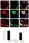
A90V TDP-43 variant results in the aberrant localization of TDP-43 in vitro
- PMID: 18505686
- PMCID: PMC2478749
- DOI: 10.1016/j.febslet.2008.05.024
A90V TDP-43 variant results in the aberrant localization of TDP-43 in vitro
Abstract
TAR DNA-binding protein-43 (TDP-43) is a highly conserved, ubiquitously expressed nuclear protein that was recently identified as the disease protein in frontotemporal lobar degeneration with ubiquitin-positive inclusions (FTLD-U) and amyotrophic lateral sclerosis (ALS). Pathogenic TDP-43 gene (TARDBP) mutations have been identified in familial ALS kindreds, and here we report a TARDBP variant (A90V) in a FTLD/ALS patient with a family history of dementia. Significantly, A90V is located between the bipartite nuclear localization signal sequence of TDP-43 and the in vitro expression of TDP-43-A90V led to its sequestration with endogenous TDP-43 as insoluble cytoplasmic aggregates. Thus, A90V may be a genetic risk factor for FTLD/ALS because it predisposes nuclear TDP-43 to redistribute to the cytoplasm and form pathological aggregates.
Figures


References
-
- Grossman M. J Int Neuropsychol Soc. 2002;8:566–83. - PubMed
-
- McKhann GM, Albert MS, Grossman M, Miller B, Dickson D, Trojanowski JQ. Arch Neurol. 2001;58:1803–9. - PubMed
-
- Neary D, et al. Neurology. 1998;51:1546–54. - PubMed
-
- Hodges JR, Davies RR, Xuereb JH, Casey B, Broe M, Bak TH, Kril JJ, Halliday GM. Ann Neurol. 2004;56:399–406. - PubMed
-
- Lomen-Hoerth C, Anderson T, Miller B. Neurology. 2002;59:1077–9. - PubMed
Publication types
MeSH terms
Substances
Grants and funding
- P30 AG010124/AG/NIA NIH HHS/United States
- R01 MH057899/MH/NIMH NIH HHS/United States
- P50 AG005142/AG/NIA NIH HHS/United States
- AG 10124/AG/NIA NIH HHS/United States
- P01 AG019724/AG/NIA NIH HHS/United States
- P30 AG010129/AG/NIA NIH HHS/United States
- AG 05142/AG/NIA NIH HHS/United States
- AG 10129/AG/NIA NIH HHS/United States
- P50 AG016574/AG/NIA NIH HHS/United States
- P01 AG017586/AG/NIA NIH HHS/United States
- P50 AG005136/AG/NIA NIH HHS/United States
- AG 16574/AG/NIA NIH HHS/United States
- MH 57899/MH/NIMH NIH HHS/United States
- AG 17586/AG/NIA NIH HHS/United States
LinkOut - more resources
Full Text Sources
Medical
Miscellaneous

