. 2008 Aug;76(8):3511-24.
doi: 10.1128/IAI.00192-08. Epub 2008 May 27.

Hookworm-induced persistent changes to the immunological environment of the lung

Affiliations

Hookworm-induced persistent changes to the immunological environment of the lung

Joshua J Reece et al. Infect Immun. 2008 Aug.

Abstract

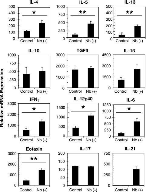
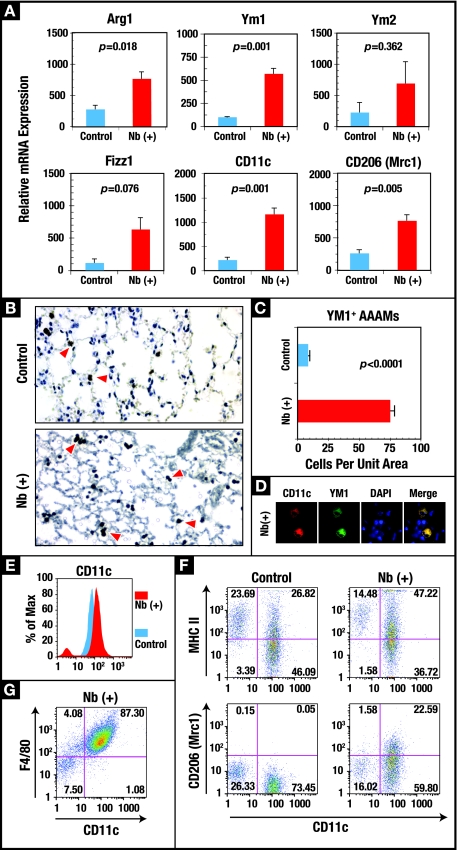
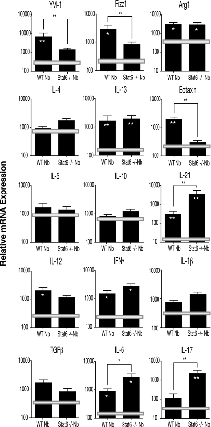
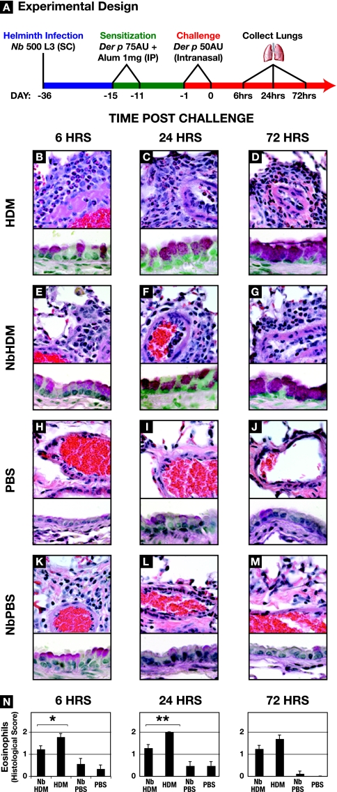
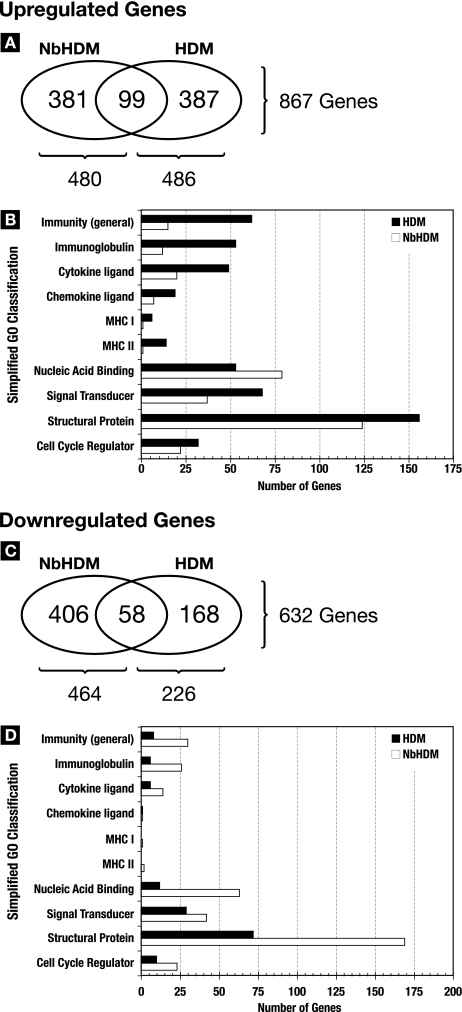
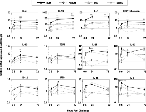
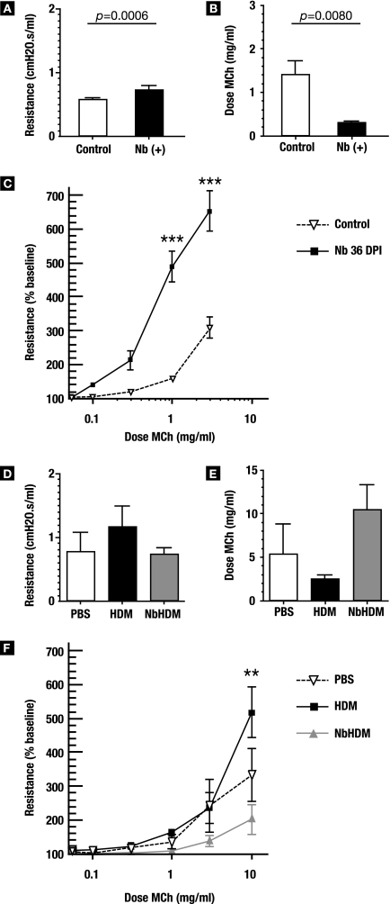
A number of important helminth parasites of humans have incorporated short-term residence in the lungs as an obligate phase of their life cycles. The significance of this transient pulmonary exposure to the infection and immunity is not clear. Employing a rodent model of infection with hookworm (Nippostrongylus brasiliensis), we characterized the long-term changes in the immunological status of the lungs induced by parasite infection. At 36 days after infection, alterations included a sustained increase in the transcription of both Th2 and Th1 cytokines as well as a significant increase in the number and frequency of alveolar macrophages displaying an alternatively activated phenotype. While N. brasiliensis did not induce alternate activation of lung macrophages in STAT6(-/-) animals, the parasite did induce a robust Th17 response in the pulmonary environment, suggesting that STAT6 signaling plays a role in modulating Th17 immunity and pathology in the lungs. In the context of the cellular and molecular changes induced by N. brasiliensis infection, there was a significant reduction in overall airway responsiveness and lung inflammation in response to allergen. In addition, the N. brasiliensis-altered pulmonary environment showed dramatic alterations in the nature and number of genes that were up- and downregulated in the lung in response to allergen challenge. The results demonstrate that even a transient exposure to a helminth parasite can effect significant and protracted changes in the immunological environment of the lung and that these complex molecular and cellular changes are likely to play a role in modulating a subsequent allergen-induced inflammatory response.


Figures

FIG. 1.
FIG. 1.
Cellular and structural changes in the lungs at 36 days post-N. brasiliensis (Nb) infection. Light microscopy of the lungs from uninfected (control) and N. brasiliensis-infected (day 36 p.i.) BALB/c mice is shown. Histological analysis illustrates focal emphysema-like lesions resulting from parasite migration (A and B) (magnification, ×10; H&E stain), peribronchial infiltration (Panels C & D, 40x magnification, H&E stain), the presence of large alveolar macrophages containing pigmented granules (E and F) (magnification, ×100; H&E stain), and an increase in the number of goblet cells (H) (magnification, ×60; PAS stain).
FIG. 2.
FIG. 2.
N. brasiliensis (Nb) induces an altered cytokine environment. Lungs from mice at 36 days p.i. were harvested and snap frozen in liquid nitrogen, RNA was extracted using Trizol, and first-strand DNA was synthesized. Real-time RT-PCR analysis of whole lungs for Th1, Th2, and regulatory cytokines, as well as eotaxin (CCL11) and IL-21, is shown. Bars represent the mean levels from five mice ± standard errors of the means. *, P < 0.05; **, P < 0.01.
FIG. 3.
FIG. 3.
The N. brasiliensis (Nb)-altered pulmonary environment is characterized by a sustained increase in CD11c+ AAMs. (A) Real-time RT-PCR was used to measure the transcript levels of the genes encoding the AAM-associated proteins ARG1, YM1, YM2, FIZZ1, CD11c, and CD206 in RNA isolated from whole lungs of uninfected control and day 36 p.i. (Nb+) BALB/c mice. Each bar represents the mean of five mice ± the standard error of the mean. (B) Lung tissue sections were immunostained with anti-YM1, and antibody binding was detected using the chromogen 3,3′-diaminobenzidine and counterstaining with H&E. YM1+ cells are shown with arrowheads. Magnification, ×10. (C) Quantification of YM1-expressing cells was calculated from six contiguous fields for triplicate mice and expressed as the number of cells per unit area. (D) Colocalization of CD11c (Texas red) and YM1 (fluorescein) in alveolar macrophages from N. brasiliensis-infected animals. Nuclei were stained with DAPI, and images were acquired using a SPOT charge-coupled device camera and software. Cells were visualized at a magnification of ×100. (E) FLOW analysis was used to measure the change in the levels of CD11c on the surface of macrophages isolated from whole-lung digests from uninfected and day 36 p.i. mice (see Materials and Methods). (F) FLOW analysis of the changes in MHC class II and CD206 (Mrc1) expression on the surface of CD11c+ cells isolated from the lungs of control and day 36 p.i. mice. (G) FLOW analysis of F4/80 expression on the CD11c+ cells from the lungs of day 36 p.i. mice. The FLOW results are representative of the results from five animals per group.
FIG. 4.
FIG. 4.
N. brasiliensis (Nb)-induced changes in the pulmonary immune environment is mediated partially by STAT6 signaling. RNA was isolated from the lungs of STAT6−/− and WT mice at day 36 post-N. brasiliensis infection, and real-time RT-PCR was used to assess the expression levels of selected cytokines and genes associated with inflammation and alternative activation of macrophages. The results were expressed in relative expression units, and each bar represents the mean threshold cycle value from five animals (± standard error of the mean). Horizontal bars represent the mean values of expression levels obtained from uninfected STAT6−/− and WT lungs. Statistical comparisons were made using a two-tailed Student t test. Significance between STAT6−/− and WT expression is designated by brackets, and significance between expression in infected and uninfected STAT6−/− or WT lungs is designated by asterisks within the bars (*, P < 0.05; **, P < 0.01).
FIG. 5.
FIG. 5.
N. brasiliensis (Nb) infection dampens allergen-induced inflammation. (A) Schedule for HDM (Der p) sensitization and challenge of uninfected or N. brasiliensis-infected BALB/c mice. (B to M) Lungs were harvested, fixed, and sectioned for histological examination at 6, 24, or 72 h after the second challenge dose of HDM. The upper portions of the panels were stained with H&E, the lower parts were stained with PAS, and both were photographed at a magnification of ×60. HDM, uninfected mice sensitized and challenged with Der p; NbHDM, day 36 p.i. mice sensitized and challenged with Der p;PBS; uninfected mice exposed to PBS; NbPBS, day 36 p.i. mice exposed to PBS. (N) Quantification of the eosinophil infiltrate. Eosinophil scoring was performed under blinded conditions from three sections representative of three different strata of the lung for each of three mice per treatment group. Scoring was as follows: 0, no eosinophils; 0.5, scattered eosinophils throughout the lung; 1.0, 10 to 40% of the perivascular infiltrate; 1.5, 50 to 60% of the perivascular infiltrate; 2.0, >70% of the perivascular infiltrate.
FIG. 6.
FIG. 6.
N. brasiliensis (Nb) modifies global gene responses to allergen challenge. Affymetrix chip-based whole-genome expression analysis was used to analyze the transcriptional responses of lungs at 6, 24, and 72 h post-allergen challenge. Changes above or below twofold were considered significant. Fold changes for the HDM and N. brasiliensis-HDM groups were calculated based on the transcriptional levels measured in age-matched uninfected and N. brasiliensis-infected (day 36 p.i.) control lungs. Expression levels are represented by the means for three mice/group/time point. (A and C) Venn diagrams showing the total numbers of genes upregulated (A) and downregulated (C) at 6, 24, and 72 h post-allergen challenge in the lungs from N. brasiliensis-HDM and HDM mice. (B and D) A simplified gene ontology breakdown of the upregulated genes (B) and downregulated genes (D) in the lungs from N. brasiliensis-HDM and HDM mice. The graphs in panels B and D include only the subset of up- and downregulated genes that were clearly classifiable into the 10 GO terms used.
FIG. 7.
FIG. 7.
N. brasiliensis (Nb) alters immune responses to allergen challenge and reduces airway resistance. Real-time RT-PCR was used to measure gene expression in the whole lungs removed from control or N. brasiliensis-infected mice at 6, 24, or 72 h postchallenge with either HDM or PBS. Fold change calculations for the N. brasiliensis-HDM and N. brasiliensis-PBS groups were made based on the gene expression levels of N. brasiliensis-infected lungs at day 36 p.i. Fold change calculations for the HDM and PBS groups were based on expression levels of age-matched uninfected controls. Points represent the mean expression levels for five mice/group/time point. Error bars represent the standard errors of the means. Statistical comparisons were generated by a two-way ANOVA followed by Bonferroni posttesting. Comparison of HDM versus N. brasiliensis-HDM: *, P < 0.05; **, P < 0.01; ***, P < 0.001. Comparison of HDM versus PBS: †, P < 0.05; ††, P < 0.01; †††, P < 0.001. Comparison of PBS versus N. brasiliensis-PBS: §, P < 0.05.
FIG. 8.
FIG. 8.
N. brasiliensis (Nb) infection alters airway responsiveness. (A to C) Groups of six day 36 p.i. and age-matched control mice were intubated and placed on a ventilator to assess their pulmonary function in vivo. (A) Baseline resistance was calculated and expressed as cm H2O·s/ml. (B) Methacholine (MCh) was then added for 10 seconds by nebulizer, and corresponding airway resistance was measured. The amount of MCh required to double the baseline resistance is shown. (C) Dose responses to MCh were normalized to a baseline resistance of 100. (D to E) N. brasiliensis-infected and control mice were sensitized and challenged with HDM as outlined in Materials and Methods. At 24 h after the second challenge, mice were sedated, intubated, and placed on a ventilator to test their responsiveness to MCh. (D) Baseline resistance in the lung before MCh challenge, expressed as cm H2O·s/ml. (E) Dose of MCh required to double the baseline resistance in the lungs. (F) Dose response to MCh challenge. Error bars represent standard errors of the means. Two-way ANOVA analysis followed by Bonferroni posttesting showed that there was a significant difference (**, P < 0.01; ***, P < 0.001).

Similar articles

Cited by

References

    1. Allen, J. E., and R. M. Maizels. 1996. Immunology of human helminth infection. Int. Arch. Allergy Immunol. 1093-10. - PubMed
    1. Anthony, R. M., J. F. Urban, Jr., F. Alem, H. A. Hamed, C. T. Rozo, J. L. Boucher, N. Van Rooijen, and W. C. Gause. 2006. Memory T(H)2 cells induce alternatively activated macrophages to mediate protection against nematode parasites. Nat. Med. 12955-960. - PMC - PubMed
    1. Araujo, M. I., A. A. Lopes, M. Medeiros, A. A. Cruz, L. Sousa-Atta, D. Sole, and E. M. Carvalho. 2000. Inverse association between skin response to aeroallergens and Schistosoma mansoni infection. Int. Arch. Allergy Immunol. 123145-148. - PubMed
    1. Arora, S., Y. Hernandez, J. R. Erb-Downward, R. A. McDonald, G. B. Toews, and G. B. Huffnagle. 2005. Role of IFN-gamma in regulating T2 immunity and the development of alternatively activated macrophages during allergic bronchopulmonary mycosis. J. Immunol. 1746346-6356. - PubMed
    1. Bashir, M. E., P. Andersen, I. J. Fuss, H. N. Shi, and C. Nagler-Anderson. 2002. An enteric helminth infection protects against an allergic response to dietary antigen. J. Immunol. 1693284-3292. - PubMed

Publication types

Associated data