. 2008 Aug;28(15):4794-804.
doi: 10.1128/MCB.00436-08. Epub 2008 May 27.

Mind bomb-1 is essential for intraembryonic hematopoiesis in the aortic endothelium and the subaortic patches

Affiliations

Mind bomb-1 is essential for intraembryonic hematopoiesis in the aortic endothelium and the subaortic patches

Mi-Jeong Yoon et al. Mol Cell Biol. 2008 Aug.

Abstract

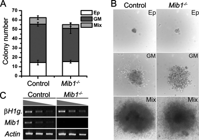
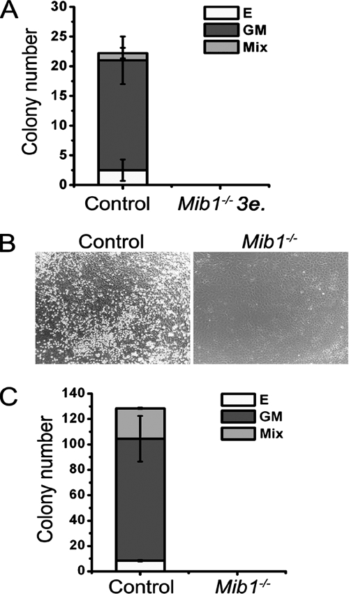
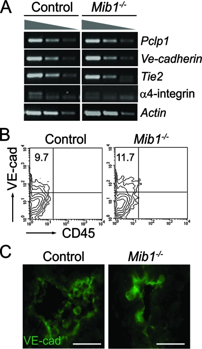
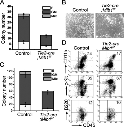
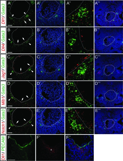
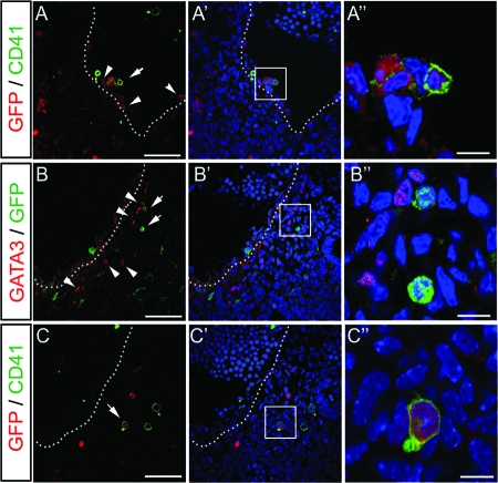
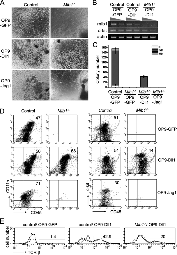
Intraembryonic hematopoiesis occurs at two different sites, the floor of the aorta and subaortic patches (SAPs) of the para-aortic splanchnopleura (P-Sp)/aorta-gonad-mesonephros (AGM) region. Notch1 and RBP-jkappa are critical for the specification of hematopoietic stem cells (HSCs) in Notch signal-receiving cells. However, the mechanism by which Notch signaling is triggered from the Notch signal-sending cells to support embryonic hematopoiesis remains to be determined. We previously reported that Mind bomb-1 (Mib1) regulates Notch ligands in the Notch signal-sending cells (B. K. Koo, M. J. Yoon, K. J. Yoon, S. K. Im, Y. Y. Kim, C. H. Kim, P. G. Suh, Y. N. Jan, and Y. Y. Kong, PLoS ONE 2:e1221, 2007). Here, we show that intraembryonic hematopoietic progenitors were absent in the P-Sp of Mib1(-/-) embryos, whereas they were partly preserved in the Tie2-cre; Mib1(f)(/f) P-Sps, suggesting that Mib1 plays a role in the endothelium and the SAPs. Interestingly, dll1 and dll4/Jag1 are expressed in the SAPs and the endothelium of the AGM, respectively, where mib1 is detected. Indeed, Notch signaling was activated in the nascent HSCs at both sites. In the P-Sp explant culture, the overexpression of Dll1 in OP9 stromal cells rescued the failed production of hematopoietic progenitors in the Mib1(-/-) P-Sp, while its activity was abolished by Mib1 knockdown. These results suggest that Mib1 is important for intraembryonic hematopoiesis not only in the aortic endothelium but also in the SAPs.


Figures

FIG. 1.
FIG. 1.
Normal hematopoiesis in the YS of Mib1−/− embryos. Cells freshly prepared from littermate control or Mib1−/− YS at E8.0 to ∼8.5 (colonies per 1 × 10⁵ cells) were cultured in semisolid medium for 7 days. (A) The colony number and type from the CFC assay were evaluated. Bars indicate means ± standard deviations of CFCs obtained from each YS in three independent experiments. Ep, primitive erythroid colony; GM, colony; Mix, mixed colony. (B) Representative colony morphologies from the CFC assay are shown. Magnification, ×100. (C) The expression levels of βH1-globin (βH1g.) and Mib1 were analyzed by semiquantitative RT-PCR of the YS cells from control and Mib1−/− embryos at E8.0.
FIG. 2.
FIG. 2.
Defective hematopoiesis in the P-Sp region of Mib1−/− embryos. (A) Direct CFC assay using fresh P-Sp cells from E9.5 littermate control and Mib1−/− embryos. Bars indicate means ± standard deviations of CFCs obtained from one control embryo and a pool of three Mib1−/− embryos (3e.) in three independent experiments. (B) P-Sp explants from E9.5 control or Mib1−/− embryos were cultured on OP9 cells for 7 days. Magnification, ×100. (C) CFC activity of the cells recovered from the P-Sp explant culture. Bars indicate means ± standard deviations of CFCs obtained from the P-Sp culture in four independent experiments. E, erythroid colony; GM, GM colony; Mix, mixed colony.
FIG. 3.
FIG. 3.
Hemogenic endothelial cells in the Mib1−/− embryos. (A) The expression of surface markers of hemogenic endothelial cells was analyzed by semiquantitative RT-PCR to compare the expression levels of the P-Sp of E9.5 control embryos to those of Mib1−/− embryos. (B) Cells from the P-Sp were analyzed by flow cytometry for the expression of VE-cadherin and CD45 gated on the Ter119 populations. A representative from three independent experiments is shown, and the percentages of cells in the upper left quadrant are indicated. (C) Immunostaining with an anti-VE-cadherin antibody on a transverse section of the aorta from E9.5 control and Mib1−/− embryos. The orientation of the aorta is dorsal (up) to ventral (down). Magnification, ×400; scale bars, 50 μm.
FIG. 4.
FIG. 4.
Preserved intraembryonic hematopoiesis in the Tie2-cre; Mib1f/f embryos. (A) Direct CFC assay using fresh P-Sp cells from E9.5 littermate control and Tie2-cre; Mib1f/f embryos. Bars represent means ± standard deviations of CFC activities obtained from control and Tie2-cre; Mib1f/f P-Sps in three independent experiments. E, erythroid colony; GM, GM colony; Mix, mixed colony. (B) P-Sp explants from E9.5 control and Tie2-cre; Mibf/f embryos were cultured on OP9 cells for 10 days. Magnification, ×100. (C) The CFC activity of cells from the Tie2-cre; Mib1f/f P-Sp explant culture (in colonies per cultured cells from P-Sp) was reduced compared to that from control P-Sp. Bars indicate the means ± standard deviations of CFCs obtained from the P-Sp culture in three independent experiments. (D) Nonadherent cells were harvested at day 12 and were analyzed for the surface expression of CD45, CD11b, c-Kit, and B220. Note that the nonadherent cells from the Tie2-cre; Mib1f/f P-Sp explants express various hematopoietic cell surface markers. Representative results from three independent experiments are presented, and the percentages of cells in the upper right quadrant are indicated.
FIG. 5.
FIG. 5.
Notch ligand expression in the AGM. (A to E‴) Fluorescent double in situ hybridization of Gata3 (in green) with either Dll1 (A), Dll4 (B), Jag1 (C), Mib1 (D), or Notch1 (E) (in red) probes on transverse sections from E10.5 wild-type AGM. (A to E) Merged images of the green and red fluorescent signals. (A′ to E′) Merged images of the green and Hoechst signals. (A″ to E″) High-magnification views of panels A to E. (A‴ to E‴) Low-magnification views of merged images of the green, red, and Hoechst signals of panels A to E. (A″)Dll1 is expressed in the mesenchyme ventral to the aorta, which was intermingled with the Gata3 transcripts. (B) Dll4 is detected mainly in the lining of the aorta, presumably the endothelium (arrow) but not in the SAPs (arrowhead). (C) Jag1 is detected in the lining of the aorta and mesenchyme (arrow) but not in the SAPs (arrowhead). (D) Mib1 is detected ubiquitously, including in aorta and SAPs (arrow). (E) Notch1 is detected in the endothelium and the SAPs (arrow). Dotted lines indicate the lining of the aorta. Arrowheads indicate the SAPs. The orientation of the aorta is dorsal (up) to ventral (down). (F to F″) Immunostaining with anti-CD31 (green) and anti-Dll1 (red) antibodies on a transverse section from E10.5 wild-type AGM. The asterisk shows the Dll1-positive patch. Scale bars, 50 μm.
FIG. 6.
FIG. 6.
Notch signaling in the AGM. (A to A″) Immunostaining with anti-CD41 (in green) and anti-GFP (in red) antibodies on transverse sections from E10.5 TNR AGM. Notch reporter activity in the TNR AGM is observed in endothelial cells and hematopoietic cells budding from the aorta wall (in red; arrowheads). In hematopoietic clusters of the aorta, Notch signaling is activated in some CD41-positive cells (arrow). (B to B″) Immunostaining with anti-GFP (in green) and anti-GATA3 (in red) antibodies on a transverse section from an E10.5 TNR embryo. GFP-positive cells (arrows) are located near the GATA3-expessing cells (arrowheads). (C to C″) Immunostaining with anti-CD41 (in green) and anti-GFP (in red) antibodies on a section sequential to that of panel B. Dotted lines indicate the lining of the aorta. (A″ to C″) High-magnification views of each of the small square regions of panels A′ to C′. Scale bars for panels A to C, 50 μm; scale bars for panels A″ to C″, 10 μm.
FIG. 7.
FIG. 7.
Restored hematopoietic activity of Mib1−/− P-Sp by OP9 cells expressing Dll1. (A) P-Sp explants from littermate control and Mib1−/− embryos were cultured on OP9-GFP, OP9-MSCV-Dll1 (OP9-Dll1), or OP9-Jag1 stromal cells for 10 days in the presence of SCF and IL-3. The white clumps are hematopoietic cells, and the dark background is the OP9 stromal cells. Black bodies are P-Sp explants. Magnification, ×40. (B) The expression levels of Mib1 and c-kit were analyzed by RT-PCR in the cells recovered from the Mib1−/− P-Sp by the OP9-MSCV-Dll1 cell cocultures. (C) The nonadherent cells from the P-Sp cocultures were harvested at day 12 and plated into the semisolid medium. Bars indicate the means ± standard deviations of CFCs obtained from the P-Sp culture in three independent experiments. E, erythroid colony; GM, GM colony; Mix, mixed colony. (D) The nonadherent cells were harvested at day 12 and were analyzed for the surface expression of CD45, CD11b, and c-Kit. Representative results from three independent experiments are presented, and the percentages of cells in the upper right quadrant are indicated. (E) P-Sp explants from control and Mib1/ embryos were cultured on OP9-GFP and OP9-MSCV-Dll1 cells in the presence of IL-7, SCF, and IL-3. The nonadherent cells were harvested at day 16 and were analyzed for the surface expression of TCR-β. The profiles indicated by the dotted lines represent cells stained without primary antibody. Percentages reflect cells considered positive.
FIG. 8.
FIG. 8.
Mib1 functions for Notch signaling in the P-Sp and OP9 stromal cells. (A) Defective Notch triggering in the Mib1/ P-Sp. Cells from littermate control (white bar) and Mib1/ (black bar) embryos were cocultured with C2C12-Notch1 cells transfected with 8× CBF-Luc and Renilla luciferase vectors. Forty-eight hours after coculture, luciferase activities were measured and normalized to Renilla luciferase activity. Data are presented as the change (n-fold) in the induction of luciferase activity relative to that of C2C12-Notch1 alone (means ± standard deviations are given). *, P < 0.002. WB w/o P-Sp, whole body without P-Sp. (B) Immunoblot of Mib1 protein in OP9-MigR1-Dll1 (Dll1) cells 36 h after microporation with control (cont si) or Mib1 (mib1 si) siRNA. Dll1 expression was not affected. (C) OP9-MigR1-Dll1 cells treated with control (cont si) or Mib1 (mib1 si) siRNA were cocultured with C2C12-Notch1 cells transfected with 8× wild-type CBF-Luc (CBF-W) or mutant CBF-Luc (CBF-M) vector. Twenty-four hours after coculture, luciferase activities were measured. **, P < 0.001. (D and E) P-Sp explants from control and Mib1−/− embryos were cultured on OP9-MigR1-Dll1 (Dll1) cells treated with either control siRNA (cont si) or Mib1 siRNA (mib1 si) in the presence of IL-7 for 12 (D) and 20 (E) days. The nonadherent cells were analyzed for the surface expression of CD45, CD11b, and c-Kit (D) and TCR-β (E). (D) Representative results from three independent experiments are presented, and the percentages of cells in the upper right quadrant are indicated. (E) The profiles indicated by the dotted lines represent cells stained without primary antibody, and percentages reflect cells considered positive. (F) Model for Mib1 function in the generation of hematopoietic progenitors of the aortic endothelium and SAPs. Mib1 is ubiquitously expressed in endothelial cells and mesenchymal cells within the P-Sp/AGM. Although Mib1 is widely expressed, Notch ligands such as Dll1, Dll4, and Jag1 are distinctively expressed. In the aortic endothelium, Mib1 regulates Dll4/Jag1 and activates Notch signaling for the specification of HSCs from hemogenic endothelium. At the SAPs, Mib1 regulates Dll1 to activate Notch signaling in pre-HSCs (the presumptive hematopoietic stem cells). Notch signaling induces the commitment to the CD41-positive nascent HSCs.

References

    1. Artavanis-Tsakonas, S., M. D. Rand, and R. J. Lake. 1999. Notch signaling: cell fate control and signal integration in development. Science 284770-776. - PubMed
    1. Bertrand, J. Y., S. Giroux, R. Golub, M. Klaine, A. Jalil, L. Boucontet, I. Godin, and A. Cumano. 2005. Characterization of purified intraembryonic hematopoietic stem cells as a tool to define their site of origin. Proc. Natl. Acad. Sci. USA 102134-139. - PMC - PubMed
    1. Brou, C., F. Logeat, N. Gupta, C. Bessia, O. LeBail, J. R. Doedens, A. Cumano, P. Roux, R. A. Black, and A. Israel. 2000. A novel proteolytic cleavage involved in Notch signaling: the role of the disintegrin-metalloprotease TACE. Mol. Cell 5207-216. - PubMed
    1. Burns, C. E., D. Traver, E. Mayhall, J. L. Shepard, and L. I. Zon. 2005. Hematopoietic stem cell fate is established by the Notch-Runx pathway. Genes Dev. 192331-2342. - PMC - PubMed
    1. Corbel, C., and J. Salaun. 2002. AlphaIIb integrin expression during development of the murine hemopoietic system. Dev. Biol. 243301-311. - PubMed

Publication types

MeSH terms

LinkOut - more resources