. 2008 Jun 3;105(22):7785-90.
doi: 10.1073/pnas.0703423105. Epub 2008 May 27.

p53 stabilization in response to DNA damage requires Akt/PKB and DNA-PK

Affiliations

p53 stabilization in response to DNA damage requires Akt/PKB and DNA-PK

Karen A Boehme et al. Proc Natl Acad Sci U S A. .

Abstract

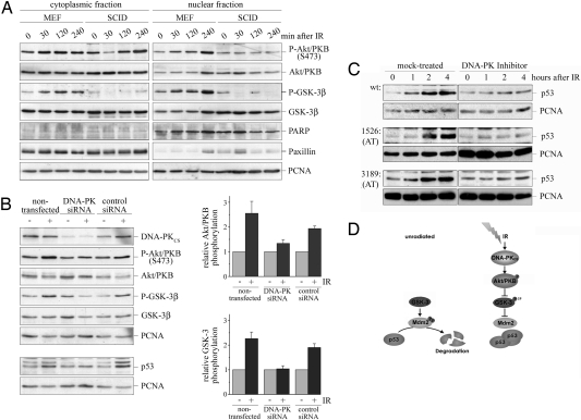
The p53 protein is one of the major tumor suppressor proteins. In response to DNA damage, p53 is prevented from degradation and accumulates to high levels. Ionizing radiation leads to hypophosphorylation of the p53 ubiquitin ligase Mdm2 at sites where phosphorylation is critical for p53 degradation and to the phosphorylation and activation of Akt/PKB, a kinase that phosphorylates and inhibits GSK-3. GSK-3, which normally phosphorylates Mdm2, is inactivated in response to ionizing radiation. We show that p53 accumulates in lymphoblasts from patients with the hereditary disorder ataxia telangiectasia in response to ionizing radiation despite the absence of a functional ATM kinase. Also, knockdown of ATR did not prevent p53 accumulation in response to ionizing radiation. Instead, p53 stabilization in response to ionizing radiation depended on the inactivation of GSK-3 and the presence of Akt/PKB. Akt/PKB is a target of DNA-PK, a kinase that is activated after ionizing radiation. Correspondingly, down-regulation of DNA-PK prevented phosphorylation of Akt/PKB and GSK-3 after ionizing radiation and strongly reduced the accumulation of p53. We therefore propose a signaling cascade for the regulation of p53 in response to ionizing radiation that involves activation of DNA-PK and Akt/PKB and inactivation of GSK-3 and Mdm2.


Conflict of interest statement

The authors declare no conflict of interest.

Figures

Fig. 1.
Fig. 1.
p53 accumulates in AT cells in response to IR. (A) Lymphoblasts from AT patients (719; 1526; and 3189), from a patient with Nijmegen breakage syndrome (NBS), and from an unaffected control were probed for ATM and for proliferating cell nuclear antigen (PCNA) for loading control. (B) Lymphoblasts from AT patients and an unaffected control were irradiated, harvested at the indicated time points, and probed for p53 and for PCNA for loading control. (C) Lymphoblasts from AT patients and an unaffected control were irradiated with the indicated doses, harvested 4 h after irradiation, and probed for p53 and PCNA. (D) Lymphoblasts from AT patients and an unaffected control were incubated in the presence of 10 μM MG132 for 4 h. Fifteen, 30, or 60 min before harvest, cells were irradiated, lysed, and probed for p53 phosphorylated at serine-15, serine-20, pan-p53, and PCNA. (E) Wild-type lymphoblasts were incubated with 10 μM MG132 for 5 h or left untreated for control. Thirty minutes before irradiation an ATM kinase inhibitor (10 μM) was added. Cells were lysed 1.5 h after irradiation and probed for p53 and PCNA. Lysates from cells that had been incubated with MG132 were also probed for p53 phosphorylated at serine-15.
Fig. 2.
Fig. 2.
Down-regulation of ATM and/or ATR does not prevent the accumulation of p53 in response to IR. U2OS cells were transfected with siRNA directed against ATM, ATR, ATM and ATR, or a nonspecific control siRNA. Ninety-six hours after transfection, cells were irradiated. Cells were harvested 4 h after irradiation. ATM, p53, and PCNA expression was determined by Western blotting. A second membrane was hybridized with antibodies directed against ATR, p53 phosphorylated at serine-15, and PCNA. Signals for p53 and PCNA were quantified. The graph shows mean values and standard deviations of three independent experiments. The relative amount of p53 in unirradiated cells was set to 1.
Fig. 3.
Fig. 3.
Inactivation of GSK-3β contributes to the accumulation of p53 in response to IR. (A) U2OS cells were incubated with 10 μM alsterpaullone for the indicated time. p53 and PCNA levels were determined by Western blotting. (B) Wild-type lymphoblasts were irradiated and harvested after the indicated time. Phosphorylated GSK-3β, total GSK-3β, and PCNA were determined by Western blotting. (C) Lymphoblasts from AT patients and unaffected controls were irradiated and harvested after the indicated times. Phosphorylated GSK-3β and PCNA were determined by Western blotting. (D) U2OS cells were transfected with Flag-tagged wild-type GSK-3β, with Flag-tagged GSK-3β-S9A, or with vector for control, selected for 6 days with neomycin, irradiated, and harvested 4 h after irradiation. p53, phosphorylated GSK-3, Flag-tagged GSK-3β, and PCNA expression was determined by Western blotting. (E) H1299 cells were transfected with 0.2 μg of a plasmid expressing p53 together with 1 μg of a plasmid expressing wild-type Mdm2 or mutant Mdm2 where serine-240, serine-254, or both were replaced with an aspartic acid or with a vector DNA for control. Forty-eight hours after transfection, cells were irradiated with 7.5 Gy. Cells were harvested 4 h after irradiation. Hdm/Mdm2, p53, and PCNA levels were determined by Western blotting. Signals for p53 and PCNA were quantified. The graph shows mean values and standard deviations of three independent experiments. The relative amount of p53 in unirradiated cells was set to 100%.
Fig. 4.
Fig. 4.
Akt/PKB phosphorylates GSK-3β in response to IR. (A) Wild-type lymphoblasts were irradiated, harvested at the indicated times, and separated into cytoplasmic and nuclear fractions. Phosphorylation of Akt/PKB at threonine-308 and serine-473, total Akt/PKB, phosphorylation of GSK-3β at serine-9, total GSK-3β, and PCNA were determined by Western blotting. (B) Lymphoblasts from AT patients and unaffected controls were irradiated and harvested after the indicated times. Phosphorylated Akt, total Akt, and PCNA were determined by Western blotting. (C) Wild-type lymphoblasts were irradiated and harvested after 4 h. Where indicated, cells were fractionated into nuclei and cytoplasm. (I) Akt/PKB was immunoprecipitated from 600 μg of lysates and incubated with 1.5 μg of GST-GSK-3β and [γ-32P]ATP. For control, total cell lysate of irradiated cells was incubated with IgG coupled to protein A Agarose (IgG) and processed as described. Samples were separated by electrophoresis and blotted. The membrane was exposed onto an x-ray film (32P-GSK-3β) before hybridization with antibodies directed against Akt/PKB and GST. (II) Lysates were separated by SDS/PAGE, blotted, and processed as described in the legend to A. Signals for phosphorylated Akt/PKB, total Akt/PKB, phosphorylated GSK-3β, and total GSK-3β were quantified, and the relative increase in phosphorylation after IR was calculated. Relative phosphorylation of Akt/PKB and GSK-3β of nonirradiated cells were set to 1. (D) U2OS cells were transfected with siRNA directed against Akt-1/PKBα, Akt-2/PKBβ, or a nonspecific control siRNA or left untransfected for control. Seventy-two hours after transfection, cells were irradiated. Cell were harvested 4 h after irradiation and probed for Akt/PKB (directed specifically against Akt-2/PKBβ in the second part of the figure), phosphorylated GSK-3β, total GSK-3β, and PCNA. A second membrane was probed for p53 and PCNA. Signals for phosphorylated GSK-3β and total GSK-3β were quantified. Relative values for GSK-3β phosphorylation of three independent experiments and standard deviations were plotted. Relative values for GSK-3β phosphorylation of unirradiated cells were set to 1.
Fig. 5.
Fig. 5.
Activation of Akt/PKB in response to IR requires DNA-PK. (A) Embryonal mouse fibroblasts (MEF) from healthy donors and from mice with SCID were irradiated, harvested at the indicated time points, fractionated into nuclei and cytoplasm, and probed for phosphorylated Akt/PKB (serine-473), total Akt/PKB, phosphorylated GSK-3β, total GSK-3β, for PARP and paxillin to monitor the quality of fractionation, and for PCNA. (B) U2OS cells were transfected with siRNA directed against the catalytic subunit of DNA-PK, a nonspecific control siRNA, or left untransfected for control. Ninety-six hours after transfection, cells were irradiated. Cells were harvested 4 h after irradiation. DNA-PK, phosphorylated Akt/PKB (serine-473), total Akt/PKB, phosphorylated GSK-3β, total GSK-3β, and PCNA were determined by Western blotting. Signals were quantified, and the relative amount of phosphorylated Akt/PKB and phosphorylated GSK-3β was calculated. Mean values and standard deviation of three independent experiments were plotted. The relative amount of phosphorylated Akt/PKB and GSK-3β was set to 1. A second membrane was hybridized with the anti-p53 antibody Ab-2 and an antibody directed against PCNA. (C) Lymphoblasts from AT patients and unaffected controls were incubated with 2 μM of an inhibitor of DNA-PK, irradiated, and harvested after the indicated time. p53 and PCNA levels were determined by Western blotting. (D) Model for the pathway for p53 control in response to IR. Activation of DNA-PK leads to the phosphorylation and activation of Akt/PKB and subsequently to the inactivation of GSK-3β. In consequence, Mdm2 is hypophosphorylated and incapable of mediating p53 degradation.

References

    1. Gasser S, Raulet D. The DNA damage response, immunity and cancer. Semin Cancer Biol. 2006;5:344–347. - PubMed
    1. Donehower LA, et al. Deficiency of p53 accelerates mammary tumorigenesis in Wnt-1 transgenic mice and promotes chromosomal instability. Genes Dev. 1995;9:882–895. - PubMed
    1. Brooks CL, Gu W. p53 ubiquitination: Mdm2 and beyond. Mol Cell. 2006;21:307–315. - PMC - PubMed
    1. Levine AJ, Hu W, Feng Z. The p53 pathway: What questions remain to be explored? Cell Death Differ. 2006;13:1027–1036. - PubMed
    1. Lavin MF, Gueven N. The complexity of p53 stabilization and activation. Cell Death Differ. 2006;6:941–950. - PubMed

Publication types

MeSH terms