Adenomatoid tumour of the liver
- PMID: 18505892
- PMCID: PMC2569191
- DOI: 10.1136/jcp.2007.054684
Adenomatoid tumour of the liver
Abstract
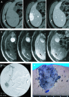
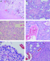
An unusual primary adenomatoid tumour arising in the normal liver is described. Hepatectomy was performed, and the patient is alive and free of disease 1 year postsurgery. Grossly, the tumour showed a haemorrhagic cut surface with numerous microcystic structures. Histological examination revealed cystic or angiomatoid spaces of various sizes lined by cuboidal, low-columnar, or flattened epithelioid cells with vacuolated cytoplasm and round to oval nuclei. The epithelioid cells were entirely supported by proliferated capillaries and arteries together with collagenous stroma. Immunohistochemical studies showed that the epithelioid cells were strongly positive for a broad spectrum of cytokeratins (AE1/AE3, CAM5.2, epithelial membrane antigen and cytokeratin 7) and mesothelial markers (calretinin, Wilms' tumour 1 and D2-40). These cells were negative for Hep par-1, carcinoembryonic antigen, neural cell adhesion molecule, CD34, CD31 and HMB45. Atypically, abundant capillaries were observed; however, the cystic proliferation of epithelioid cells with vacuoles and immunohistochemical profile of the epithelioid element were consistent with hepatic adenomatoid tumour.
Conflict of interest statement
Figures


References
-
- Stephenson TJ, Mills PM. Adenomatoid tumours: an immunohistochemical and ultrastructural appraisal of their histogenesis. J Pathol 1986;148:327–35 - PubMed
-
- de Klerk DP, Nime F. Adenomatoid tumors (mesothelioma) of testicular and paratesticular tissue. Urology 1975;6:635–41 - PubMed
-
- Ghossain MA, Chucrallah A, Kanso H, et al. Multilocular adenomatoid tumor of the ovary: ultrasonographic findings. J Clin Ultrasound 2005;33:233–6 - PubMed
-
- Isotalo PA, Keeney GL, Sebo TJ, et al. Adenomatoid tumor of the adrenal gland: a clinicopathologic study of five cases and review of the literature. Am J Surg Pathol 2003;27:969–77 - PubMed
-
- Kaplan MA, Tazelaar HD, Hayashi T, et al. Adenomatoid tumors of the pleura. Am J Surg Pathol 1996;20:1219–23 - PubMed
Publication types
MeSH terms
Substances
LinkOut - more resources
Full Text Sources
Medical
Research Materials
Miscellaneous
