Review
. 2008 May;6(5):675-84.
doi: 10.1158/1541-7786.MCR-07-2180.

Signal transducer and activator of transcription-3: a molecular hub for signaling pathways in gliomas

Affiliations
Review

Signal transducer and activator of transcription-3: a molecular hub for signaling pathways in gliomas

Emily C Brantley et al. Mol Cancer Res. 2008 May.

Abstract

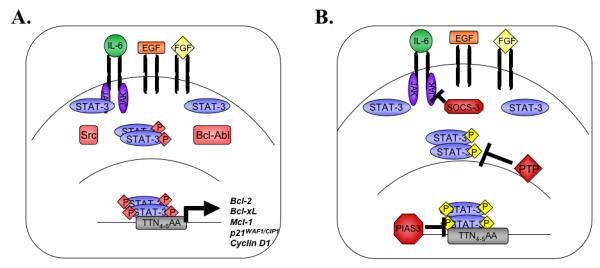
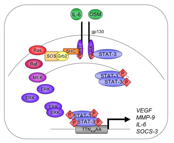
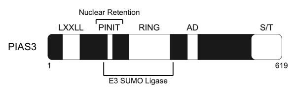
Glioblastoma is the most common and severe primary brain tumor in adults. Its aggressive and infiltrative nature renders the current therapeutics of surgical resection, radiation, and chemotherapy relatively ineffective. Accordingly, recent research has focused on the elucidation of various signal transduction pathways in glioblastoma, particularly aberrant activation. This review focuses on the signal transducer and activator of transcription-3 (STAT-3) signal transduction pathway in the context of this devastating tumor. STAT-3 is aberrantly activated in human glioblastoma tissues, and this activation is implicated in controlling critical cellular events thought to be involved in gliomagenesis, such as cell cycle progression, apoptosis, angiogenesis, and immune evasion. There are no reports of gain-of-function mutations in glioblastoma; rather, the activation of STAT-3 is thought to be a consequence of either dysregulation of upstream kinases or loss of endogenous inhibitors. This review provides detailed insight into the multiple mechanisms of STAT-3 activation in glioblastoma, as well as describing endogenous and chemical inhibitors of this pathway and their clinical significance. In glioblastoma, STAT-3 acts a molecular hub to link extracellular signals to transcriptional control of proliferation, cell cycle progression, and immune evasion. Because STAT-3 plays this central role in glioblastoma signal transduction, it has significant potential as a therapeutic target.


Figures

Figure 1
Figure 1. The STAT-3 Signaling Pathway
A. Cytokines such as IL-6, or growth factors including EGF and FGF, initiate STAT-3 signal transduction when they bind to their receptors and activate intracellular kinases. JAK proteins or receptor tyrosine kinases recruit inactive STAT-3 monomers and phosphorylate them on tyrosine 705. STAT-3 is also activated directly through interaction with the oncogenic kinases Src and Bcl-Abl. Tyrosine phosphorylated STAT-3 dimerizes and translocates to the nucleus, where STAT-3 transcriptional regulation is further modulated by serine phosphorylation. Active STAT-3 dimers bind to consensus sequences in the promoters of genes that regulate cell growth and anti-apoptotic behavior. B. Multiple STAT-3 endogenous inhibitors attenuate STAT-3 signaling. SOCS-3 inhibits JAK activation and subsequent signal transduction in the cytoplasm, while PIAS3 inhibits STAT-3 DNA binding in the nucleus. Protein tyrosine phosphatases such as SHP-1 and SHP-2 dephosphorylate active STAT-3 complexes.
Figure 2
Figure 2. IL-6 Cytokine Signaling Pathways
Members of the IL-6 cytokine family bind to a common receptor subunit, gp130, which either homo-or hetero-dimerizes upon ligand binding. JAK proteins are auto- and trans-phosphorylated and phosphorylate one of four tyrosine residues on the intracellular portion of the gp130 subunit. JAK protein activation results in downstream signal transduction through both the MAPK and STAT-3 pathways. Receptor interactions with the adaptor proteins SHC and Grb2 activate the MEK-ERK pathway. Alternatively, STAT-3 monomers are recruited to the receptor, become tyrosine phosphorylated and dimerize through their SH2 domains. STAT-3 dimers translocate to the nucleus and bind to consensus sequences in the promoters of genes including VEGF, MMP-9, IL-6, and SOCS-3.
Figure 3
Figure 3. Structural Domains of the PIAS3 Protein
PIAS3 is a 619 amino acid protein that shares 40% homology to the other PIAS proteins. PIAS3 contain an N-terminal LXXLL motif, a PINIT motif, which is involved in nuclear retention, a zinc ring finger domain (RING), a C-terminal acidic domain (AD) and a serine/threonine (S/T) rich domain. The PINIT motif and the RING finger domain are involved in the E3 SUMO ligase activity exhibited by PIAS3.
Figure 4
Figure 4. Inhibition of STAT-3 Signal Transduction
A variety of endogenous and pharmacologic inhibitors can attenuate STAT-3 signaling. SOCS-3, PIAS3, and various protein tyrosine phosphatases (PTP), inhibit STAT-3 activity endogenously. STAT-3 specific siRNA degrades STAT-3 mRNA. Pharmacologic inhibition of JAK activity by AG490 and WP1066 dampens the signals that result in STAT-3 activation. Attenuation of FGF signaling by dobesilate also inhibits STAT-3 mediated gene expression by attenuating kinase signals upstream of STAT-3 activation.

References

    1. Ohgaki H, Kleihues P. Epidemiology and etiology of gliomas. Acta Neuropathol (Berl) 2005;109:93–108. - PubMed
    1. Louis DN, Ohgaki H, Wiestler OD, et al. The 2007 WHO classification of tumours of the central nervous system. Acta Neuropathol (Berl) 2007;114:97–109. - PMC - PubMed
    1. Schwartzbaum JA, Fisher JL, Aldape KD, Wrensch M. Epidemiology and molecular pathology of glioma. Nat Clin Pract Neurol. 2006;2:494–503. - PubMed
    1. Stupp R, Mason WP, van den Bent MJ, et al. Radiotherapy plus concomitant and adjuvant temozolomide for glioblastoma. N Engl J Med. 2005;352:987–96. - PubMed
    1. Rao RD, James CD. Altered molecular pathways in gliomas: an overview of clinically relevant issues. Semin Oncol. 2004;31:595–604. - PubMed

Publication types