. 2008 May;6(5):795-807.
doi: 10.1158/1541-7786.MCR-07-2097.

Neutral sphingomyelinase-3 is a DNA damage and nongenotoxic stress-regulated gene that is deregulated in human malignancies

Affiliations

Neutral sphingomyelinase-3 is a DNA damage and nongenotoxic stress-regulated gene that is deregulated in human malignancies

Chad A Corcoran et al. Mol Cancer Res. 2008 May.

Abstract

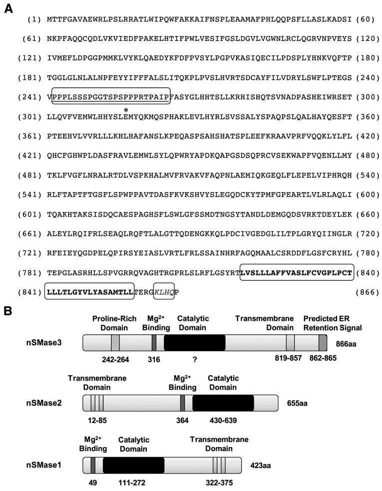
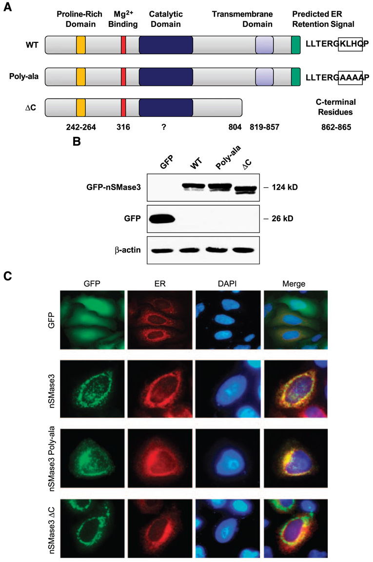
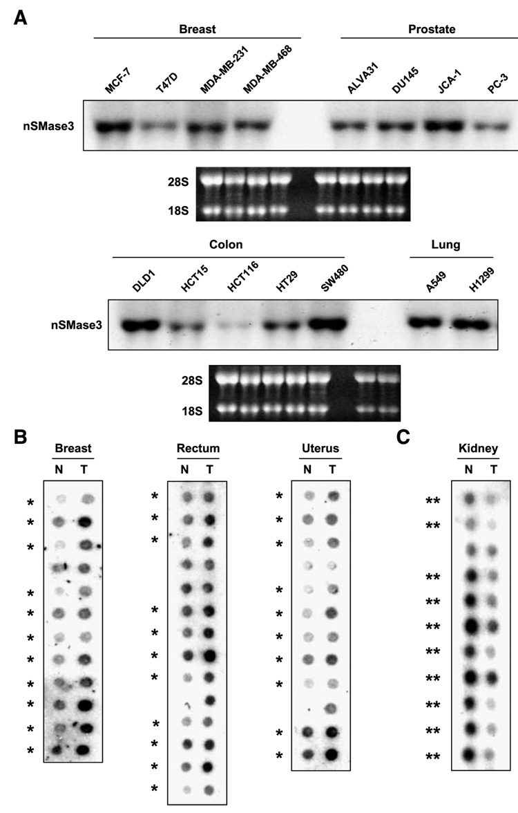
In this study, we report the characterization of a novel genotoxic and nongenotoxic stress-regulated gene that we had previously named as SKNY. Our results indicate that SKNY encodes the recently identified neutral sphingomyelinase-3 (nSMase3; hereafter SKNY is referred to as nSMase3). Examination of nSMase3 subcellular distribution reveals nSMase3 to localize to the endoplasmic reticulum (ER), and deletion of a COOH-terminal region containing its putative transmembrane domain and ER targeting signal partly alters its compartmentalization to the ER. Treatment with genotoxic Adriamycin and nongenotoxic tumor necrosis factor-alpha up-regulates endogenous nSMase3 expression, albeit with different kinetics. Tumor necrosis factor-alpha up-regulates nSMase3 expression within 2 h that lasts beyond 24 h and declines to control levels by 36 h. Adriamycin up-regulation of nSMase3 is transient, occurs within 30 min, and declines to control levels by 120 min. Prolonged treatment with Adriamycin by 24 h and beyond, however, causes a down-regulation in nSMase3 expression. Activation of wild-type p53 also down-regulates nSMase3 expression, suggesting that DNA damage-mediated nSMase3 down-regulation seems to occur partly through the tumor suppressor p53. Overexpression of exogenous nSMase3 sensitizes cells to Adriamycin-induced cell killing, a finding consistent with the proposed proapoptotic role of nSMase enzymes and nSMase-generated ceramide. We further investigated nSMase3 expression in various human malignancies and found its expression to be deregulated in several types of primary tumors when compared with their matching normal tissues. Collectively, our results have identified nSMase3 to be an important molecule that is linked to tumorigenesis and cellular stress response.


Conflict of interest statement

Disclosure of Potential Conflicts of Interest

No potential conflicts of interest were disclosed.

Figures

FIGURE 1
FIGURE 1. nSMase3 protein sequence and schematic illustration of nSMase functional motifs
A. Amino acid sequences of human nSMase3. The predicted proline-rich (242–264) region is underlined. The putative glutamic acid (Glu316) involved in Mg2+ binding is denoted by an asterisk. The predicted transmembrane-spanning region (819–857) is in bold letters and the predicted KKXX-like ER retention signal KLHQ (862–865) is italicized. B. Schematic illustration of putative nSMase3 domains; schematic illustration of known nSMase1 and nSMase2 domains is also shown for comparison.
FIGURE 2
FIGURE 2. Subcellular distribution of GFP-tagged nSMase3
A. Schematic illustration of full-length and mutation/deletion constructs of nSMase3. Numbers represent the amino acid residues corresponding to putative functional domains. Predicted KKXX-like signal (KLHQ) in WT nSMase3 and those residues changed to alanine in nSMase3 (Poly-ala) are boxed. B. Western blotting was done using an anti-GFP antibody to detect GFP and GFP-nSMase3 expression. The same blot was then probed for β-actin to show equal protein loading. C. HeLa cells were cotransfected with expression vectors carrying GFP or GFP-nSMase3 with pDsRed2-ER using Lipofectamine 2000. Cells were washed with PBS, fixed with 4% paraformaldehyde, and incubated with 4′,6-diamidino-2-phenylindole (DAPI) to stain nuclei. Cells were analyzed with an Olympus AX70 fluorescent microscope and photographs were captured using a digital camera. Photomicrographs of GFP-transfected cells are not to scale with those transfected with GFPnSMase3 expression vectors.
FIGURE 3
FIGURE 3. TNF-α and DNA-damaging agents regulate nSMase3 mRNA expression
A. A representative Northern blot showing the effect TNF-α treatment on endogenous nSMase3 mRNA levels in HT29 cells. Cells were either left untreated or treated with 50 ng/mL TNF-α for 2, 4, 8, 12, 24, 36, or 48 h. COX-2, cyclooxygenase-2. B. A representative Northern blot showing the effect of short-term Adriamycin treatment on endogenous nSMase3 mRNA levels in RKO cells. Cells were either left untreated or treated with 1 µmol/L Adriamycin for 30, 60, 90, or 120 min. C. A graphic representation of nSMase3 levels depicted in B. Relative nSMase3 mRNA levels were determined by normalizing nSMase3 mRNA to 18S rRNA. D. A representative Northern blot showing the effect of long-term Adriamycin treatment on endogenous nSMase3 mRNA levels. Cells were either left untreated or treated with 0.50 µmol/L (RKO), 1 µmol/L (DLD1), or 5 µmol/L (HT29) of Adriamycin for 12, 24, 36, or 48 h. A, B, and D. Total RNA was harvested and then subjected to Northern blot analysis as described in Materials and Methods. The blots were sequentially probed with 32P-labeled nSMase3 and cyclooxygenase-2 or p21 probes; ethidium bromide staining of the 28S and 18S rRNA is also shown to illustrate loading and RNA integrity.
FIGURE 4
FIGURE 4. nSMase3 is down-regulated by p53
A representative Northern blot showing the effect of inducible WT p53 on endogenous nSMase3 mRNA levels in DLD1 cells. Cells grown in the presence (p53 repressed) or absence (p53 induced) of 40 ng/mL doxycycline (Dox) for indicated periods of time were harvested, RNA extracted, and subjected to Northern blot analysis as described in Materials and Methods. The blot was sequentially probed with 32P-labeled nSMase3 and p21WAF1 probes; ethidium bromide staining of the 28S and 18S rRNA is also shown for RNA loading and integrity.
FIGURE 5
FIGURE 5. nSMase3 sensitizes cells to DNA damage-induced apoptosis
A. A representative Western blot showing the stable expression of exogenous Myc-tagged nSMase3 in DLD1 or RKO cells. Pooled vector (pcDNA3.1-Myc) transfectants and isolated single clones stably expressing exogenous nSMase3 (pcDNA3.1-Myc-nSMase3) were harvested and subjected to SDS-PAGE. Western blotting was done using an anti-Myc antibody. The same blot was then probed for β-actin to show equal protein loading. B. Membrane fractions from RKO and DLD1 vector and nSMase3 stable cell lines in two independent stable clones named 55 and 61, and 7 and 35, respectively. C and D. Vector and nSMase3 stable cell lines were treated with 20 nmol/L (RKO) or 40 nmol/L (DLD1) of Adriamycin for 24 or 48 h and clonogenic survivals were assessed as described in Materials and Methods. Representative plates from an RKO (C) and DLD1 (D) experiment are shown. E. DLD1 vector (○) and nSMase3-clone 7 (●) and nSMase3-clone 35 (■) stable transfectants were treated continuously for 48, 72, or 96 h with 200 nmol/L Adriamycin and 3-(4,5-dimethylthiazol-2-yl)-2,5-diphenyltetrazolium bromide cell viability assays were done as described in Materials and Methods. The experiment was done in triplicate. Points, average of triplicates; bars, SE.
FIGURE 6
FIGURE 6. nSMase3 is differentially expressed in matched normal and tumor samples
A. Representative Northern blots showing nSMase3 expression in breast, prostate, colon, and lung cancer cell lines. The blots were probed with 32P-labeled nSMase3 probes; ethidium bromide staining of the 28S and 18S rRNA is also shown to illustrate loading and RNA integrity. JCA-1 cells were previously considered as prostate cancer cells; now, they are believed to represent bladder cancer. B and C. Cancer profiling arrays containing normalized cDNA from tumor (T) and corresponding normal (N) tissue samples from individual patients were probed with nSMase3 probe as described in Materials and Methods. *, matched pairs showing higher nSMase3 mRNA levels in tumor versus matched normal tissue samples; **, matched pairs showing lower nSMase3 mRNA levels in tumor versus matched normal tissue samples.

References

    1. Zheng W, Kollmeyer J, Symolon H, et al. Ceramides and other bioactive sphingolipid backbones in health and disease: lipidomic analysis, metabolism and roles in membrane structure, dynamics, signaling and autophagy. Biochim Biophys Acta. 2006;1758:1864–1884. - PubMed
    1. Gulbins E, Li PL. Physiological and pathophysiological aspects of ceramide. Am J Physiol Regul Integr Comp Physiol. 2006;290:R11–R26. - PubMed
    1. Ogretmen B, Hannun YA. Biologically active sphingolipids in cancer pathogenesis and treatment. Nat Rev Cancer. 2004;4:604–616. - PubMed
    1. Taha TA, Mullen TD, Obeid LM. A house divided: ceramide, sphingosine, and sphingosine-1-phosphate in programmed cell death. Biochim Biophys Acta. 2006;1758:2027–2036. - PMC - PubMed
    1. Mathias S, Pena LA, Kolesnick RN. Signal transduction of stress via ceramide. Biochem J. 1998;335:465–480. - PMC - PubMed

Publication types