. 2008 Jun 3;98(11):1803-9.
doi: 10.1038/sj.bjc.6604375. Epub 2008 May 27.

Cisplatin alters nitric oxide synthase levels in human ovarian cancer cells: involvement in p53 regulation and cisplatin resistance

Affiliations

Cisplatin alters nitric oxide synthase levels in human ovarian cancer cells: involvement in p53 regulation and cisplatin resistance

E L Leung et al. Br J Cancer. .

Abstract

The present study determines if (1) basal protein levels of nitric oxide (NO) synthases (eNOS, iNOS, and nNOS) are different in cisplatin-sensitive (OV2008) and counterpart cisplatin-resistant (C13(*)) human ovarian cancer cells, (2) cisplatin alters NOS levels, (3) NO donor causes apoptosis and p53 upregulation, (4) NO donor sensitizes C13(*) cells to cisplatin via p53 upregulation (determined by p53 siRNA gene-knockdown), and (5) inhibition of endogenous NOS alters cisplatin-induced apoptosis. Basal iNOS levels were higher in OV2008 cells than in C13(*) cells. Cisplatin upregulated iNOS, but dramatically reduced eNOS and nNOS, in OV2008 cells only. Failure of cisplatin to upregulate iNOS and downregulate eNOS/nNOS in cisplatin-resistant C13(*) cells may be an aetiological factor in the development of resistance. The NO donor S-nitroso-N-acetylpenicillamine (SNAP) increased p53 protein levels and induced apoptosis in both cell types, and enhanced cisplatin-induced apoptosis in C13(*) cells in a p53-dependent manner (i.e., enhancement blocked by p53 siRNA). Specific iNOS inhibitor 1400W partially blocked cisplatin-induced apoptosis in OV2008 cells. In cisplatin-resistant C13(*) cells, blocking all NOSs with N(G)-amino-L-arginine dramatically changed these cells from cisplatin-resistant to cisplatin-sensitive, greatly potentiating cisplatin-induced apoptosis. The data suggest important roles for the three NOSs in regulating chemoresistance to cisplatin in ovarian cancer cells.


Figures

Figure 1
Figure 1
Protein levels of iNOS, eNOS, and nNOS in cisplatin-sensitive OV2008 and in cisplatin-resistant C13* cells were determined using western blot analysis. (A) Basal iNOS levels were significantly higher in OV2008 cells compared with C13* cells. Cisplatin (CDDP, 10 μM, 24 h) significantly upregulated iNOS in OV2008, but not in C13* cells. (B and C) Basal levels of eNOS and nNOS were significantly lower in OV2008 cells compared with C13* cells. Cisplatin (5 and 10 μM, 24 h) significantly downregulated both eNOS and nNOS in OV2008, but not in C13*, cells. The bar graphs show mean±s.e.m. of protein levels of five independent experiments. *P<0.05, compared with control (no cisplatin). **P<0.01, compared with control (no cisplatin). #P<0.05, comparing basal levels in control OV2008 and in C13* cells. ##P<0.01, comparing basal levels in control OV2008 and in C13* cells.
Figure 2
Figure 2
(A) OV2008 cells were cultured with SNAP (0, 100, 200, 400 μM) for 24 h, followed by 5 μM cisplatin (CDDP) for another 24 h. p53 protein content was assessed by western blot analysis. Results were normalised for protein loading by re-probing with anti-GAPDH antibody. Western blots are representative of four independent experiments. Graphs show mean±s.e.m. of protein contents of four independent experiments. S-nitroso-N-acetylpenicillamine alone (400 μM, *P<0.05) increased basal p53 content, but decreased cisplatin-induced upregulation of p53 (**P<0.01) in chemosensitive OV2008 cells. S-nitroso-N-acetylpenicillamine (400 μM) alone significantly (**P<0.01) increased apoptosis. However, when used in combination with cisplatin, SNAP did not further enhance cisplatin-induced apoptosis. (B) C13* cells were cultured with SNAP (0, 50, 100, 200, 400 μM) for 24 h, followed by 10 μM cisplatin for another 24 h. Western blots are representative of five independent experiments. Graphs show mean±s.e.m. of p53 protein content and apoptosis of five independent experiments. S-nitroso-N-acetylpenicillamine (200 and 400 μM) alone significantly (***P<0.001) upregulated p53 levels. With cisplatin co-treatment, SNAP at 50, 100, 200, and 400 μM significantly (**P<0.01, ***P<0.001) increased p53 levels above control. p53 upregulation induced by SNAP plus cisplatin was significantly (##P<0.01) larger than with SNAP alone. S-nitroso-N-acetylpenicillamine (200 and 400 μM) alone significantly (**P<0.01) induced apoptosis and sensitised chemoresistant C13* cells to cisplatin-induced apoptosis (SNAP at 200 μM, #P<0.05).
Figure 3
Figure 3
(A) Both OV2008 and C13* cells were transfected with p53-specific siRNA (50 nM; scrambled sequence as control) for 24 h, and subsequently treated with 400 μM SNAP for another 24 h. Western blots are representative of four independent experiments. Graphs show mean±s.e.m. of four independent experiments. Western blots show that p53 was successfully knocked down. *P<0.05, ***P<0.001, compared with SNAP treatment alone. (B) C13* cells were transfected with p53-specific siRNA (50 nM; scrambled sequence as control) for 24 h, and subsequently treated with 200 μM SNAP before cisplatin (CDDP, 10 μM) treatments for another 24 h. Western blots are representative of four independent experiments and graphs show mean±s.e.m. from four independent experiments. **P<0.01, compared with SNAP alone, ***P<0.001, compared with SNAP plus cisplatin (without siRNA).
Figure 4
Figure 4
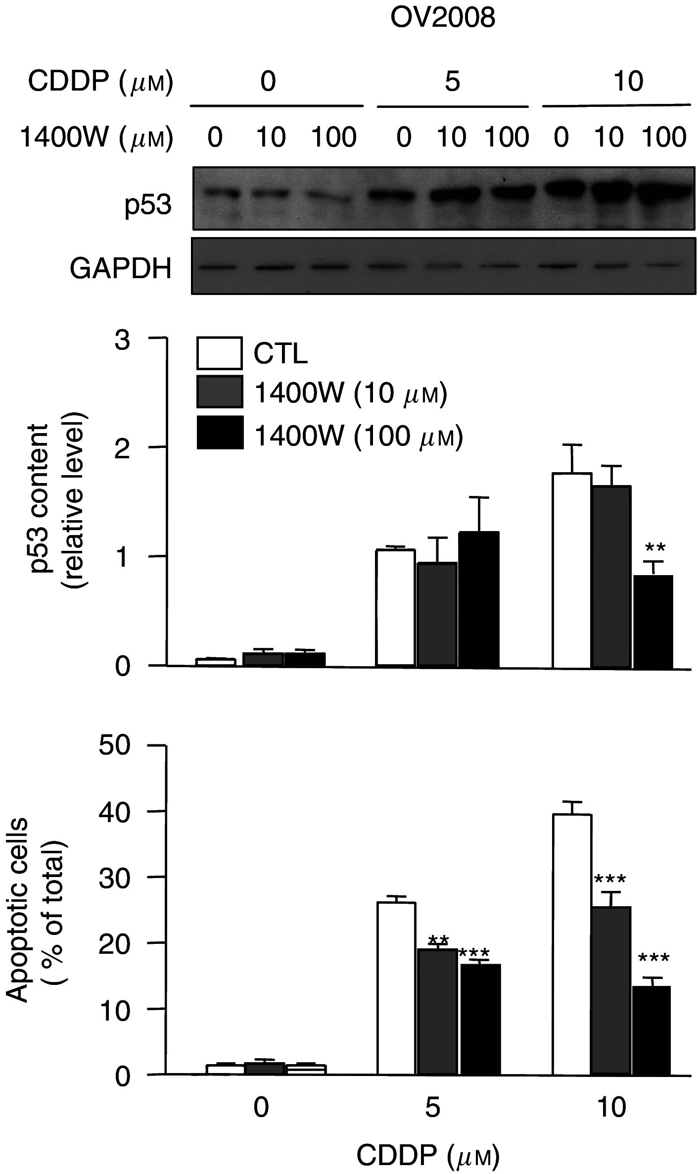
OV2008 cells were cultured with 1400W (0, 10, 100 μM) for 24 h and further treated with cisplatin (CDDP; 0, 5, 10 μM) for another 24 h. Protein content of p53 was assessed by western blot analysis and apoptosis by Hoechst staining. Western blots are representative of four independent experiments. Graphs show mean±s.e.m. of four independent experiments. **P<0.01, ***P<0.001, compared with control.
Figure 5
Figure 5
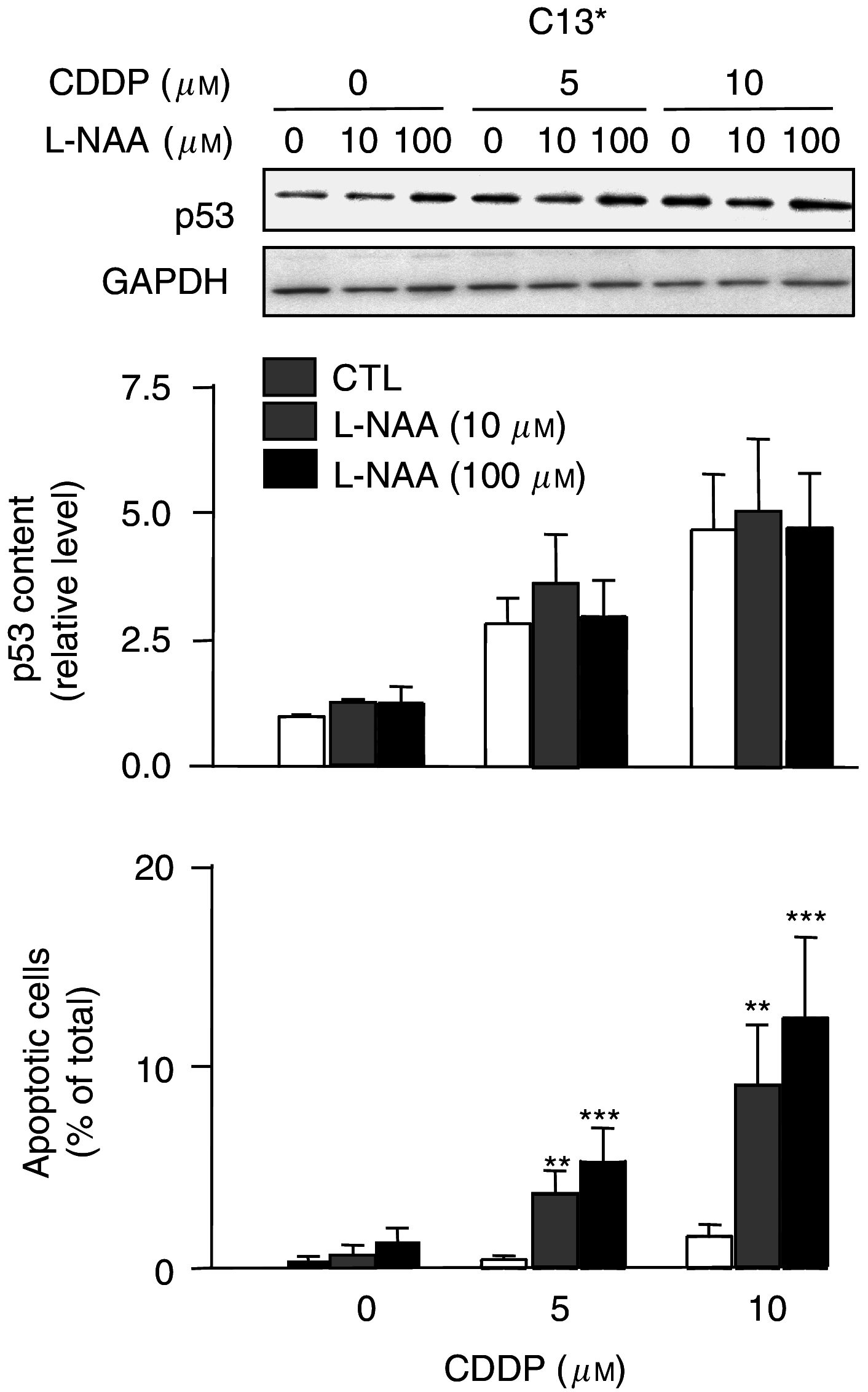
Inhibition of all NOSs using NG-amino-L-arginine (L-NAA; 0, 10, 100 μM) for 24 h, followed by cisplatin (0, 5, 10 μM) for additional 24 h. Western blots are representative of four independent experiments and graphs shows mean±s.e.m. from four independent experiments. **P<0.01, ***P<0.001.

References

    1. Ambs S, Hussain SP, Harris CC (1997) Interactive effects of nitric oxide and the p53 tumor suppressor gene in carcinogenesis and tumor progression. FASEB J 11: 443–448 - PubMed
    1. Bellamy TC, Griffiths C, Garthwaite J (2002) Differential sensitivity of guanylyl cyclase and mitochondrial respiration to nitric oxide measured using clamped concentrations. J Biol Chem 277: 31801–31807 - PubMed
    1. Chan SL, Fiscus RR (2003) Guanylyl cyclase inhibitors NS2028 and ODQ and protein kinase G (PKG) inhibitor KT5823 trigger apoptotic DNA fragmentation in immortalized uterine epithelial cells: anti-apoptotic effects of basal cGMP/PKG. Mol Hum Reprod 9: 775–783 - PubMed
    1. Cheng Chew SB, Leung PY, Fiscus RR (2003) Preincubation with atrial natriuretic peptide protects NG108-15 cells against the toxic/proapoptotic effects of the nitric oxide donor S-nitroso-N-acetylpenicillamine. Histochem Cell Biol 120: 163–171 - PubMed
    1. Fiscus RR (2002) Involvement of cyclic GMP and protein kinase G in the regulation of apoptosis and survival in neural cells. Neurosignals 11: 175–190 - PubMed

Publication types

MeSH terms