

19F NMR studies of the D-galactose chemosensory receptor. 1. Sugar binding yields a global structural change
- PMID: 1850619
- PMCID: PMC2904549
- DOI: 10.1021/bi00231a021
19F NMR studies of the D-galactose chemosensory receptor. 1. Sugar binding yields a global structural change
Abstract
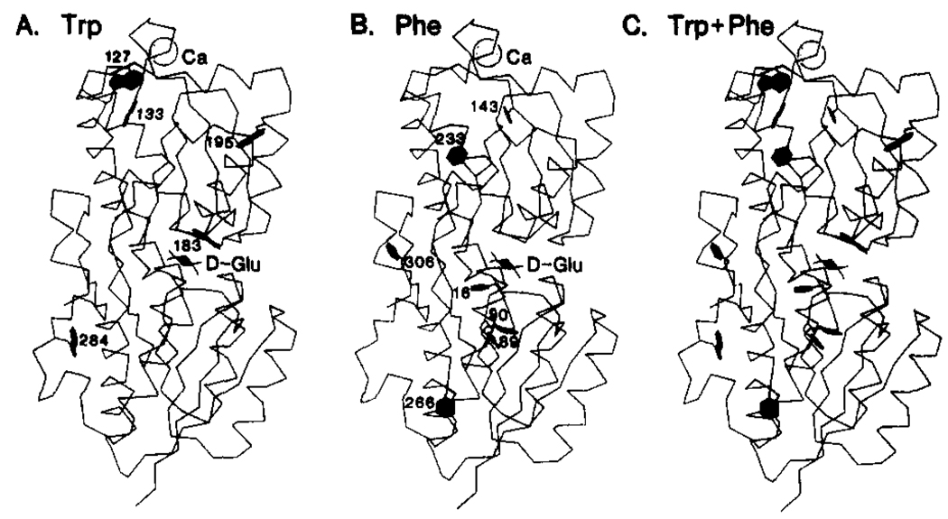
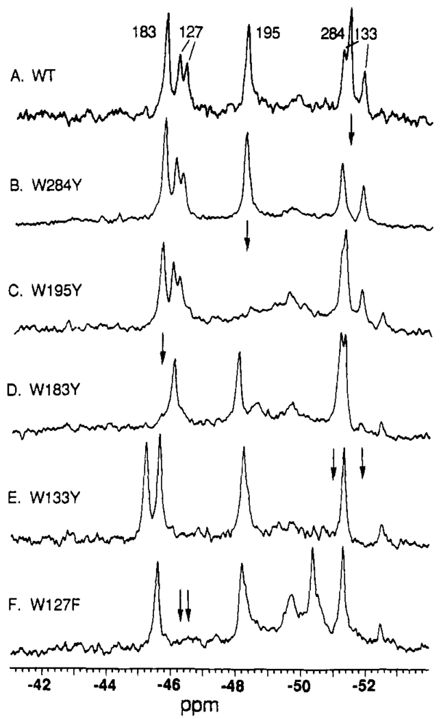
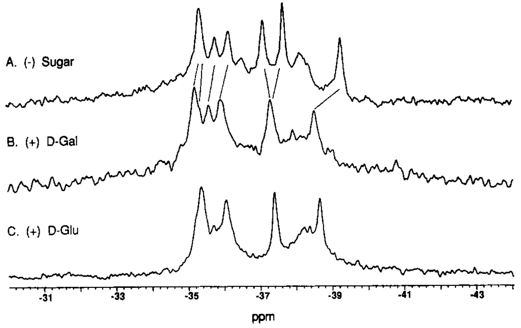
The Escherichia coli D-galactose and D-glucose receptor is an aqueous sugar-binding protein and the first component in the distinct chemosensory and transport pathways for these sugars. Activation of the receptor occurs when the sugar binds and induces a conformational change, which in turn enables docking to specific membrane proteins. Only the structure of the activated receptor containing bound D-glucose is known. To investigate the sugar-induced structural change, we have used 19F NMR to probe 12 sites widely distributed in the receptor molecule. Five sites are tryptophan positions probed by incorporation of 5-fluorotryptophan; the resulting 19F NMR resonances were assigned by site-directed mutagenesis. The other seven sites are phenylalanine positions probed by incorporation of 3-fluorophenylalanine. Sugar binding to the substrate binding cleft was observed to trigger a global structural change detected via 19F NMR frequency shifts at 10 of the 12 labeled sites. Two of the altered sites lie in the substrate binding cleft in van der Waals contact with the bound sugar molecule. The other eight altered sites, specifically two tryptophans and six phenylalanines distributed equally between the two receptor domains, are distant from the cleft and therefore experience allosteric structural changes upon sugar binding. The results are consistent with a model in which multiple secondary structural elements, known to extend between the substrate cleft and the protein surface, undergo shifts in their average positions upon sugar binding to the cleft. Such structural coupling provides a mechanism by which sugar binding to the substrate cleft can cause structural changes at one or more docking sites on the receptor surface.
Figures


Similar articles
-
19F NMR studies of the D-galactose chemosensory receptor. 2. Ca(II) binding yields a local structural change.Biochemistry. 1991 Apr 30;30(17):4257-61. doi: 10.1021/bi00231a022. Biochemistry. 1991. PMID: 1850620 Free PMC article.
-
Open conformation of a substrate-binding cleft: 19F NMR studies of cleft angle in the D-galactose chemosensory receptor.Biochemistry. 1991 Jul 2;30(26):6484-90. doi: 10.1021/bi00240a019. Biochemistry. 1991. PMID: 1647202 Free PMC article.
-
19F NMR spectroscopy of [6-19F]tryptophan-labeled Escherichia coli dihydrofolate reductase: equilibrium folding and ligand binding studies.Biochemistry. 1994 May 10;33(18):5502-9. doi: 10.1021/bi00184a019. Biochemistry. 1994. PMID: 8180172
-
Use of 19F NMR to probe protein structure and conformational changes.Annu Rev Biophys Biomol Struct. 1996;25:163-95. doi: 10.1146/annurev.bb.25.060196.001115. Annu Rev Biophys Biomol Struct. 1996. PMID: 8800468 Free PMC article. Review.
-
Detection of site-specific binding and co-binding of ligands to macromolecules using 19F NMR.Life Sci. 1991;48(13):1227-40. doi: 10.1016/0024-3205(91)90517-f. Life Sci. 1991. PMID: 2002752 Review.
Cited by
-
'Clickable lectins': bioorthogonal reactive handles facilitate the directed conjugation of lectins in a modular fashion.Interface Focus. 2019 Apr 6;9(2):20180072. doi: 10.1098/rsfs.2018.0072. Epub 2019 Feb 15. Interface Focus. 2019. PMID: 30842873 Free PMC article.
-
Activation of the phosphosignaling protein CheY. I. Analysis of the phosphorylated conformation by 19F NMR and protein engineering.J Biol Chem. 1993 Jun 25;268(18):13081-8. J Biol Chem. 1993. PMID: 8514749 Free PMC article.
-
Chemical shifts and three-dimensional protein structures.J Biomol NMR. 1995 Apr;5(3):217-25. doi: 10.1007/BF00211749. J Biomol NMR. 1995. PMID: 7787420 Review.
-
Evidence that both ligand binding and covalent adaptation drive a two-state equilibrium in the aspartate receptor signaling complex.J Gen Physiol. 2001 Dec;118(6):693-710. doi: 10.1085/jgp.118.6.693. J Gen Physiol. 2001. PMID: 11723162 Free PMC article.
-
Alternative modes of binding of proteins with tandem SH2 domains.Protein Sci. 2000 Mar;9(3):570-9. doi: 10.1110/ps.9.3.570. Protein Sci. 2000. PMID: 10752619 Free PMC article.
References
-
- Ames GF-L. Annu. Rev. Biochem. 1986;55:397–425. - PubMed
-
- Bennett WS, Huber R. Crit. Rev. Biochem. 1984;15:291–384. - PubMed
-
- Boos W, Gordon AS, Hall RE, Price DH. J. Biol. Chem. 1972;247:917–924. - PubMed
-
- Bourret R, Hess J, Borkovitch K, Simon MI. J. Biol. Chem. 1989;264:7085–7088. - PubMed
-
- Bowie JU, Reidharr-Olson JF, Lim WA, Sauer RT. Science. 1990;247:1306–1310. - PubMed
Publication types
MeSH terms
Substances
Grants and funding
LinkOut - more resources
Full Text Sources
Other Literature Sources