

Molecular markers of radiation-related normal tissue toxicity
- PMID: 18506399
- PMCID: PMC2800946
- DOI: 10.1007/s10555-008-9138-7
Molecular markers of radiation-related normal tissue toxicity
Abstract
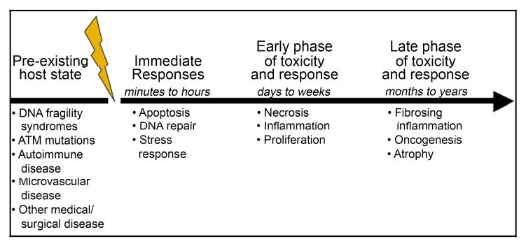
Over the past five decades, those interested in markers of radiation effect have focused primarily on tumor response. More recently, however, the view has broadened to include irradiated normal tissues-markers that predict unusual risk of side-effects, prognosticate during the prodromal and therapeutic phases, diagnose a particular toxicity as radiation-related, and, in the case of bioterror, allow for tissue-specific biodosimetry. Currently, there are few clinically useful radiation-related biomarkers. Notably, levels of some hormones such as thyroid-stimulating hormone (TSH) have been used successfully as markers of dysfunction, indicative of the need for replacement therapy, and for prevention of cancers. The most promising macromolecular markers are cytokines: TGFbeta, IL-1, IL-6, and TNFalpha being lead molecules in this class as both markers and targets for therapy. Genomics and proteomics are still in nascent stages and are actively being studied and developed.
Figures
References
-
- Radford IR. Evidence for a general relationship between the induced level of DNA double-strand breakage and cell-killing after X-irradiation of mammalian cells. Int J Radiat Biol. 1986;49(4):611–620. - PubMed
-
- Chapman JD, Allalunis-Turner MJ. Cellular and molecular targets in normal tissue radiation injury. In: Gutin PH, Liebel SA, Sheline GE, editors. Radiation injury to the nervous system. New York: Raven Press; 1991. pp. 1–16.
-
- Anscher MS, Crocker IR, Jirtle RL. Transforming growth factor-beta 1 expression in irradiated liver. Radiat Res. 1990;122(1):77–85. - PubMed
-
- Finkelstein JN, Johnston CJ, Baggs R, Rubin P. Early alterations in extracellular matrix and transforming growth factor beta gene expression in mouse lung indicative of late radiation fibrosis. Int J Radiat Oncol Biol Phys. 1994;28(3):621–631. - PubMed
-
- Piguet PF. Is “tumor necrosis factor” the major effector of pulmonary fibrosis? Eur Cytokine Netw. 1990;1(4):257–258. - PubMed
Publication types
MeSH terms
Substances
Grants and funding
LinkOut - more resources
Full Text Sources
Other Literature Sources
Medical
Miscellaneous
